Japanese
English
- 有料閲覧
- Abstract 文献概要
- 1ページ目 Look Inside
- サイト内被引用 Cited by
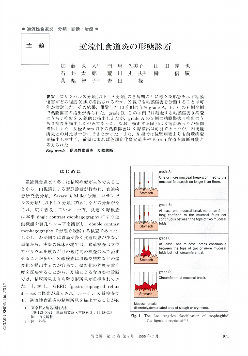
要旨 ロサンゼルス分類(以下LA分類)の各病期ごとに様々な形態を示す粘膜傷害がどの程度X線で描出されるのか,X線でも粘膜傷害を分類することは可能か検討した.その結果,供覧した10症例のうちgrade A,B,Cの6例全例で粘膜傷害の描出が得られた.grade B,Cの4例では縦走する粘膜傷害9病変のうち7病変をX線的に描出しえたが,grade Aの2例の粘膜傷害4病変のうち2病変を描出したのみであった.なお,横走する陥凹は3病変あったが全例描出しえた.長径5mm以下の粘膜傷害はX線描出は可能であったが,内視鏡所見との対比は十分にできなかった.また,X線では後壁病変よりも前壁病変が描出しやすく,前壁に限れば色調変化型食道炎やBarrett食道も診断可能と考えられた.
Radiological features of 10 patients with reflux esophagitis comfirmed by endoscopy were studied. Esophagogram by double contrast method was carried out. The morphologic findings of “mucosal breaks” diagnosed by endoscopy appeared on esophagograms as longitudinal or transverse depressed lesions surrounded by ill-defined marginal elevations. Seven of ten mucosal breaks 5 mm or more in length were easily demonstrated, and all of the three continuous mucosal breaks detected were clearly demonstrated as tranverse depressed lesions with luminal narrowing. However, only two of four mucosal breaks less than 5 mm in length were correlated with endoscopic findings. To delineate such small lesions is not difficult, but it is difficult to correlate them with endoscopic findings. The findings of depressed lesions at the anterior wall of the esophagus were demonstrated more larily than those at the posterior wall. Double contrast esophagography in the recumbent position can demonstrate fine mucosal patterns, such as Barrett's esophagus and mild esophagitis showing only discoloration without mucosal breaks.
Copyright © 1999, Igaku-Shoin Ltd. All rights reserved.