Japanese
English
- 有料閲覧
PPV購入案内
1,320円 (1,200円+税10%) こちらは、555ページから559ページの文献です。購入には医書.jp本会員登録が必要です。
会員登録完了後に改めて上記「購入サイトへ」を選択してください。
または「購入サイトへ」選択後に「購入ログイン」、「新規会員登録」の順に進んでください。
会員登録について詳しくはをご確認ください。
- Abstract 文献概要
- 1ページ目 Look Inside
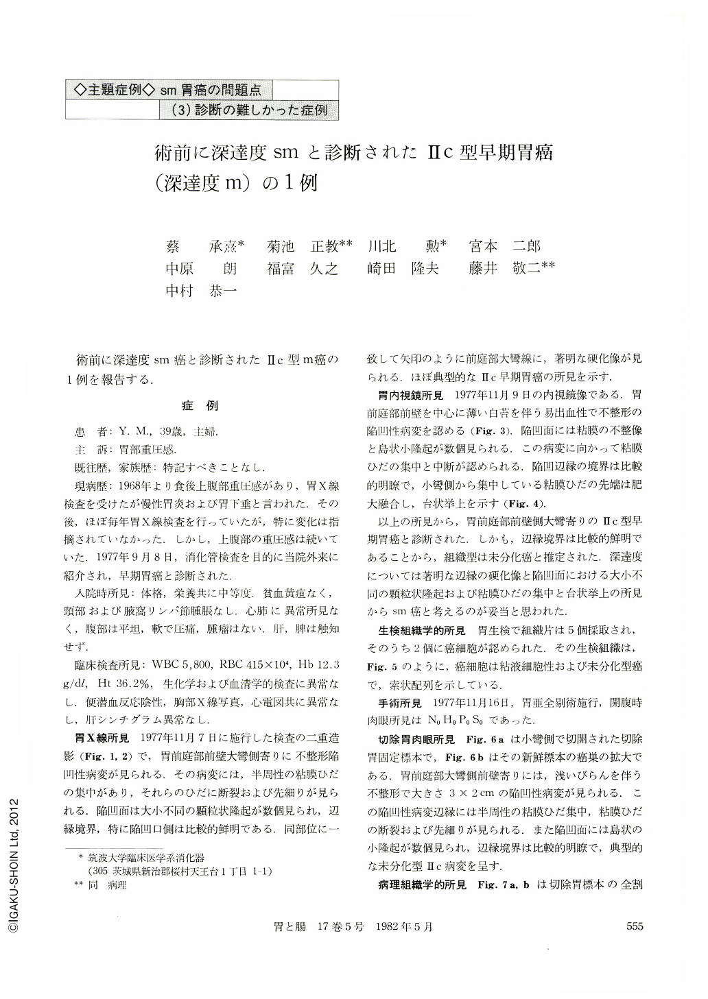
術前に深達度sm癌と診断されたⅡc型m癌の1例を報告する.
症例
患 者:Y.M.,39歳,主婦
主 訴:胃部重圧感
既往歴,家族歴:特記すべきことなし. 現病歴:1968年より食後上腹部重圧感があり,胃X線検査を受けたが慢性胃炎および胃下垂と言われた.その後,ほぼ毎年胃X線検査を行っていたが,特に変化は指摘されていなかった.しかし,上腹部の重圧感は続いていた.1977年9月8日,消化管検査を目的に当院外来に紹介され,早期胃癌と診断された.
A 39-year-old housewife was admitted to our hospital with chief complaint of sensation of abdominal fullness after meals. The double contrast picture of the stomach showed a shallow depressive lesion accompanying with convergence and interruption of rugal folds on the anterior wall of the antrum near the greater curvature. Irregularity and rigidity were also noted on the corresponding area (Fig. 1, 2). The endoscopic picture revealed an irregularly shaped ulcer on the anterior wall of the antrum with a rough ulcer base and a raised platform-like margin. Based on these findings, a Type Ⅱc infiltrating the submucosa was considered.
Biopsy materials revealed undifferentiated carcinoma showing trabecular pattern (Fig. 5). In the resected stomach, the lesion showed a typical Type Ⅱc of undifferentiated carcinoma, measuring about 4×3 cm in dimensions and located in the anterior wall of the antrum near the greater curvature (Fig. 6, 7). The Ⅱc lesion was diagnosed histologically as undifferentiated carcinoma showing trabecular arrangement and signet-ring cell pattern, and limited to the mucosa (Fig. 9). The Ⅱc lesion was accompanied with an ulcer of Ul-Ⅱ, in which extensive fibrosis of the submucosa was noted (Fig. 8).
Rigidity of early cancerous lesion has been generally mentioned as a radiological sign of submucosal invasion of cancer cells. Although a double contrast picture showed radiologically and endoscopically rigidity in this case, cancer was histologically limited to the mucosa. Radiological rigidity of the gastric wall was attributable to extensive fibrosis of the submucosa. Consequently, radiological rigidity is not always correspond to submucosal invasion of cancer cells when there is ulcerative lesion in early gastric cancer.
Copyright © 1982, Igaku-Shoin Ltd. All rights reserved.