Japanese
English
- 有料閲覧
- Abstract 文献概要
- 1ページ目 Look Inside
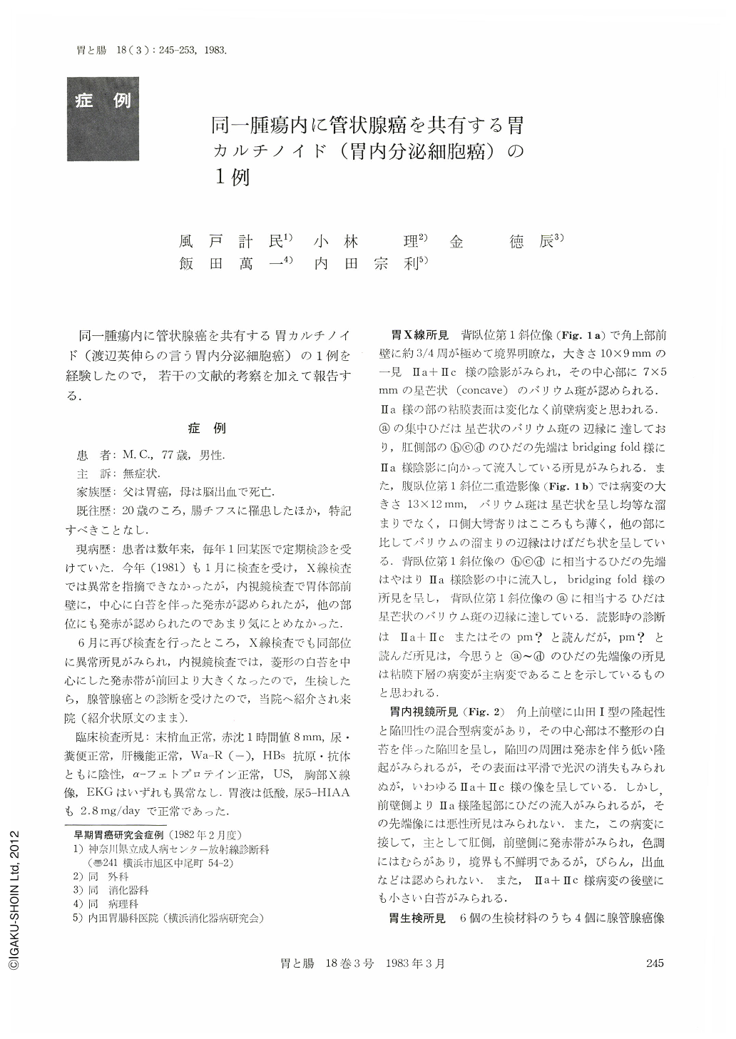
同一腫瘍内に管状腺癌を共有る胃カルチノイド(渡辺英伸らの言う胃内分泌細胞癌)の1例を経験したので,若干の文献的考察を加えて報告する.
The patient was a 77-year-old asymptomatic man. Upper gastrointestinal series and endoscopy disclosed Ⅱa+Ⅱc like lesion (7×7mm) at the anterior wall and just above the angulus, therefore six biopsies were taken from the lesion. Four out of the six biopsies showed tubular adenocarcinoma and three of them demonstrated proliferation of solid nests consisting of densely stained small cells as well as acinar structure―suggesting carcinoid. Histologically, the depth of carcinoid invasion was sm and that of the tubular adenocarcinoid was m, and moderate lymphatic metastasis were also noted.
Recently Pearse (1970) reported that gastric carcinoid belongs to APUD oma and much attention has been paid for it. Watanabe, et al. classified cell tumor the following two categories: namely classical carcinoid and endocrine cell carcinoma, then discussed the latter's histoembryology at the 41th Japan Cancer Society Meeting. He reported that two out of 13 examined gastric endocrine cell carcinoma had coexisted tubular adenocarcinoma in the same tumor, two cases had coexisted signet-ring cell carcinoma, three cases had the both types of the carcinoma and the remaining six cases were inadequately studied.
Our case was a rare one of gastric carcinoid with small coexisting tubular adenocarcinoma in the same tumor which is classified as gastric endocrine cell carcinoma by Watanabe's classification or as type D by Soga's classification. So far, Murakami et al reported 114 cases of Japanese gastric carcinoid (The Japanese Journal of Clinical and Experimental Medicine 58: 851), but we feel that more cases will be detected from now on by utilizing silver staining and EM study.

Copyright © 1983, Igaku-Shoin Ltd. All rights reserved.


