Japanese
English
- 有料閲覧
PPV購入案内
1,320円 (1,200円+税10%) こちらは、937ページから941ページの文献です。購入には医書.jp本会員登録が必要です。
会員登録完了後に改めて上記「購入サイトへ」を選択してください。
または「購入サイトへ」選択後に「購入ログイン」、「新規会員登録」の順に進んでください。
会員登録について詳しくはをご確認ください。
- Abstract 文献概要
- 1ページ目 Look Inside
脳卒中発作覚醒後より上腹部愁訴が続き,その後の検査で胃潰瘍の存在が確認され,以後加療を続けて来た患者を検査する機会があり,胃角部前壁にⅡc+Ⅲタイプの微小胃癌を発見した.今日前壁病変の診断は比較的容易となって来たが,卒中後遺症患者の場合,その取り扱いに留意せねばならぬ点がある.本例も左半身不随があるためどうしてもX線にて病変の性状を描写することができず,内視鏡検査および生検にて確診しえた例である.
Ⅰ.症例
患 者:T. N. 64歳 男
主 訴:胃精検の希望.
家族歴:癌死はない.
既往歴:10年前より高血圧症で時々医治を求めていた他,特別なことはない.ことに胃腸に関する愁訴は全くなかった.
In recent years lesions on the anterior wall of the stomach in otherwise healthy persons are being more accurately diagnosed, but in patients in postapoplecatal stage these lesions are often difficult to examine because of hemiplegia or other bodily conditions. The case here described seems to corroborate the necessity of careful examination of the stomach in such physically hampered patients.
Case : a 64-year-old man. Two weeks after he had been attacked with apoplexia in September 1969, he began to suffer from increasing gastric distress. One year later, he was examined by x-ray because of full sensation of the stomach, and was diagnosed as gastric ulcer on the posterior wall of the corpus associated with another small excavated lesion on the anterior wall at the level of the angle. Although ulcer on the posterior wall of the corpus was gone after three months' hospitalization, the other lesion of the anterior wall of the angle remained unchanged. For yet another five months he was treated as intractable ulcer, but the lesion persisted. Accordingly he was referred to our hospital in April 1971.
Present status : left hemiplegia and scoliosis. The abdomen was of normal consistency and no tumor was palpated. Laboratory reports were all within normal limits.
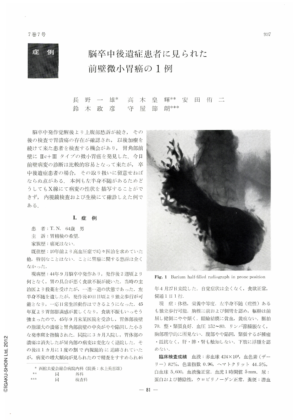
X-ray examination : Because of his stooped posture, it was hard delineate his stomach, but in a double contrast picture of the anterior wall in prone position was seen a localized, shallow barium fleck on the anterior wall of the angle with rugal convergence toward it. These ceased folds were clubby at the tips.
Endoscopic findings : A small, oval, and slightly irregularly excavated lesion was noted on the antorior wall of the angulus region. It was covered with white coat, and the surrounding mucosa was strongly reddened. Some converging folds were irregularly interrupted at the reddened margins. Of nine mucosal fragments obtained by biopsy under direct vision, two proved to be malignant.
Subtotal gastrectomy was performed (S0 N0 H0 P0).
Findings of the resected stomach : An irregularly shaped shallow depression, measuring 6×8 mm, was seen in the center of the anterior wall of the angulus region. A very small ulcer was observed in the center. The whole lesion even including the marginal reddened areas was less than 10 mm in diameter. It was judged as very minute early gastric cancer of type Ⅱc+Ⅲ. Histologically, it proved to be adenocarcinoma tubulare localized within the mucosal layer.
Copyright © 1972, Igaku-Shoin Ltd. All rights reserved.