Japanese
English
- 有料閲覧
- Abstract 文献概要
- 1ページ目 Look Inside
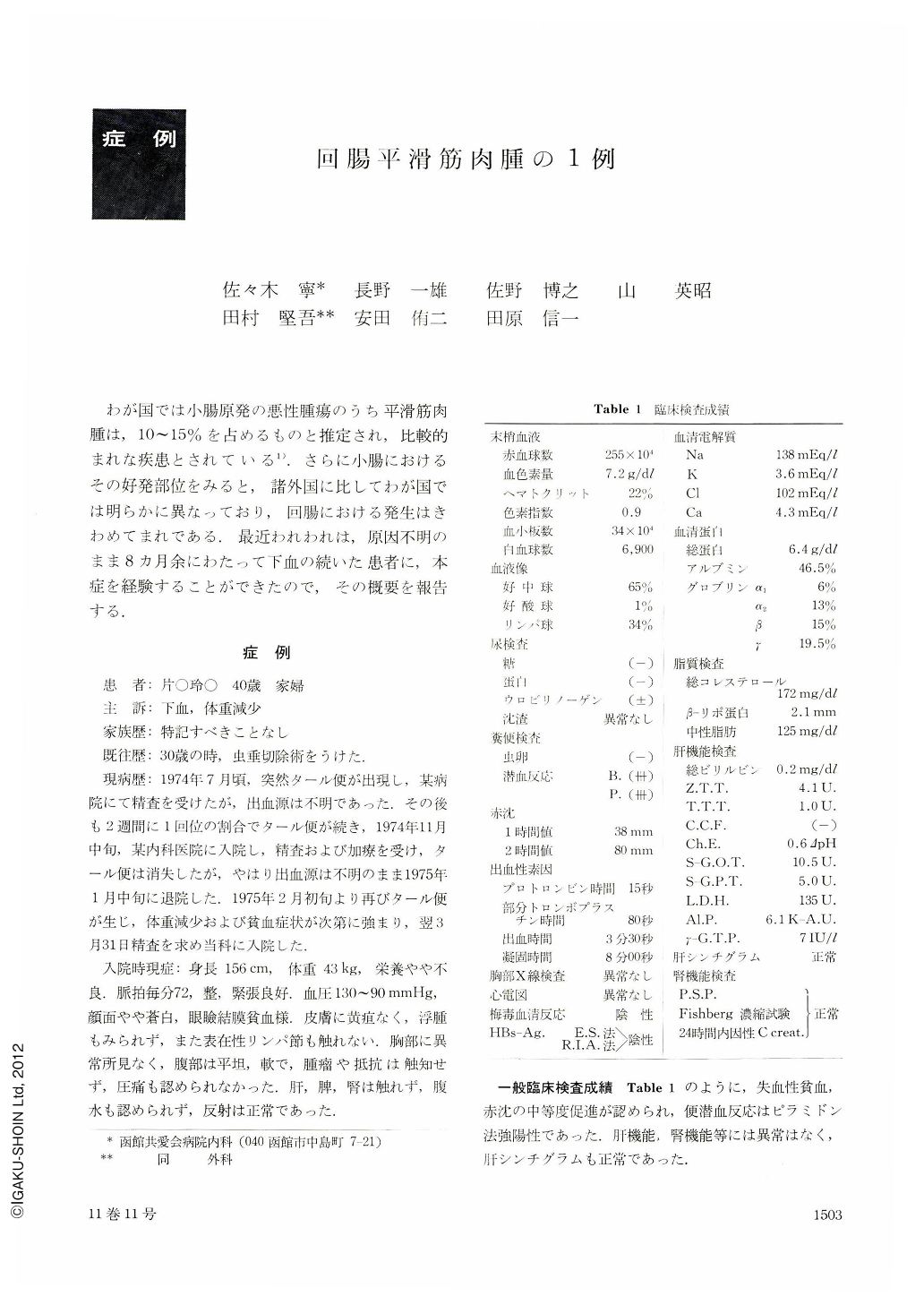
わが国では小腸原発の悪性腫瘍のうち平滑筋肉腫は,10~15%を占めるものと推定され,比較的まれな疾患とされている1).さらに小腸におけるその好発部位をみると,諸外国に比してわが国では明らかに異なっており,回腸における発生はきわめてまれである.最近われわれは,原因不明のまま8カ月余にわたって下血の続いた患者に,本症を経験することができたので,その概要を報告する.
The incidence of primary malignant tumors of the small intestine is extremely low. In Europe and America, these malignancies are said to account for about 6.0% of all malignant tumors of the gastrointestinal tracts, and, in Japan, they are said to account for 1.5 to 3.1% of all malignant tumors of the intestine. Leiomyosarcoma is a relatively rare disease, which is estimated as accounting for 10 to 15% of them in Japan. Recently, the authors encountered a case of leiomyosarcoma originating from the ileum, and the findings are summarized here.
It was a case in a 40 year old housewife. She had tarry stools about once every two weeks since about July 1974. She was admitted to the authers' hospital for examination in detail on March 31, 1975. Clinical examinations on admission revealed no abnormalities other than anemia (hemoglobin 7.2 g per dl). Neither routine X-ray examination of the upper and lower alimentary tract nor endoscopy disclosed any abnormalities, so that we were led to a suspicion of a lesion in the small intestine. Barium meal examination showed a large filling defect in the ileum due to an elevated lesion. This elevated lesion had a coarse, nodular surface, with a barium spot of irregular contour at its top. The ileum was slightly dilated on its oral side. She was operated on under a diagnosis of a malignant tumor of the ileum. The tumor was located about 60 cm on the oral side of Bauhin's valve, and there was no metastasis to the surrounding organs and the regional lymph nodes. The resected specimen measured 10×8×6 cm. It had grown both inside and outside of the intestinal canal, with its center at the part of the ileum to which mesentery is attached; the central part of the tumor had formed a hemorrhagic necrotic focus, and the mucosal surface was ulcerated. Histopathologically, spindle cells had proliferated fascicularly, and their nuclei were of spindle shape, and hyperchromatic. The mitosis was observed at a rate of one in every several 200×magnification fields. The lesion was diagnosed as a leiomyosarcoma with low malignancy. The patient has followed a favorable postoperative course without any sign of metastasis or recurrence for 11 months since the operation.
To the best of our knowledge a total of 25 cases of leiomyosarcoma of the ileum have been reported in Japan from 1932 to 1975 and this case is the 26th one in our country.
Copyright © 1976, Igaku-Shoin Ltd. All rights reserved.