Japanese
English
- 有料閲覧
- Abstract 文献概要
- 1ページ目 Look Inside
臨床的に胃粘膜の巨大皺襞は,大別してただひだが大きいだけのもの,始めてMénétrierが唱えたような概念のもの,及び悪性腫瘍に関連のあるものに分けられる1).このうち良性の巨大皺襞を青山2),高木3)は巨大皺襞症と呼び,わが国では広く用いられるようになってきた.しかし,これが以前より学者によって種々の名前4)5)で呼ばれ,その診断基準や病理組織像についても随分相違する所に問題があった.Ménétrier病をめぐる従来の主な混乱は,巨大皺襞と本病との混同で,X線検査で認めるgiant rugae内視鏡検査で認めるgiant foldを組織像の裏付けもなくhypertrophic gastritisと呼んだものがあることもあって,両者を等しいとする見解が生じたものであり,現在Ménétrier病は巨大皺襞を示す疾患の一つであるが,原著者6)がもっとも力を入れている組織学的所見が具っていることが本症の条件となっている7)8).筆者らは最近,広汎な胃polyposisを伴った胃巨大皺襞症を経験した.血清蛋白動態は正常であったが,切除標本の検討などからこれがMénétrierのPolyadénomes polypeuxとPolyadénomes en nappeの併存例に相当することを知った.さらに本例にはS字状結腸,直腸にpolyposisを合併しており,Ménétrier病の場合の下部消化管病変については意外に報告が乏しいので多少の文献的考察を加え報告した.
Giant hypertrophy of the gastric mucosa with diffuse polyposis, coexistence of themselves were very rarely, associated with polyposis of the colon, was recently encountered and surgically treated. Surprisingly seldom has been the description of lesions of the lower digestive canal in literature of the Ménétrier's disease up to now, some reference has also been made both to this problem points and to its literature in general.
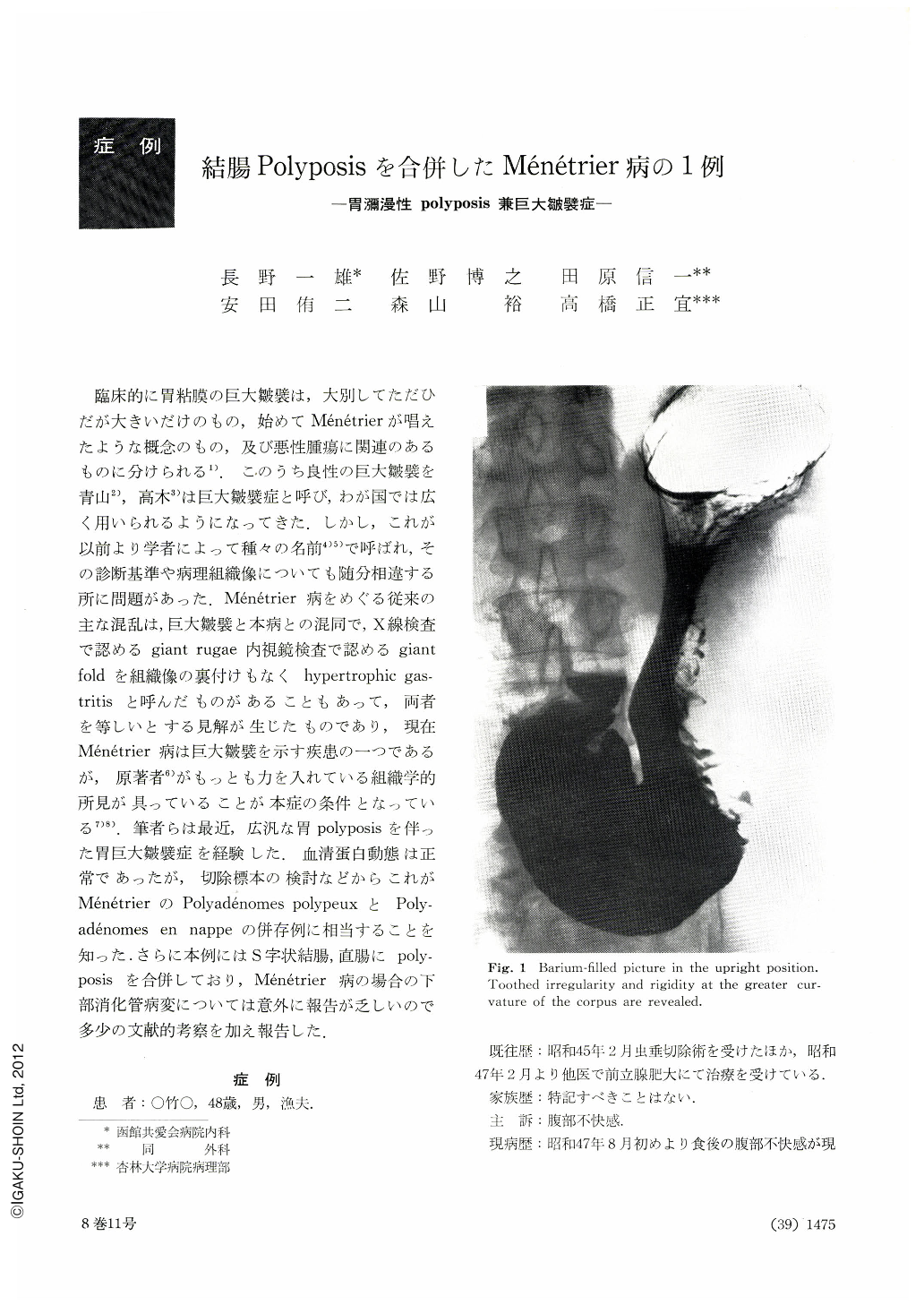
The patient, a man 48 years of age, was seen first on September 1972 because of unpleasant sensation around the navel directly after meals and he was admitted to the author's hospital for thorough examination. Except for slight tenderness around the appendectomical scar and in the lower right quadrant, physical examination was non-contributory. No pigmentation, no edema and no nail or bone dystrophy was recognized. Gastrointestinal series disclosed irregular, tortuous, highly-thickened mucosal rugae at the corpus and numerous sessile small polypoid lesions in the pyloric antrum, which also extend into the angular region of lesser curvature and the cardia. Barium enema study showed many flat small polypoid defects, especially in the rectum and sigmoid. Gastrofibrescopic study revealed numerous polypoid lesions, which were belonged to type Ⅰ or Ⅱ type as designed by Yamada and noted no reddness or depression on their surfaces, in the pyloric antrum, angular region and cardia, and coarse tortuous giant folds looked like cerebral convolutions. Hypersecretion of gastric mucus was noticed. Colonofibrescopic study revealed also numerous sessile polypoid lesions in the rectum and sigmoid. These surface were same as surrounding colonal mucosa. Biopsy specimens taken from polyposis and giant folds of the stomach and polyposis of the colon disclosed Group type Ⅰ. Gastric juice showed normal acidity and plasma protein level was 6.8 g/dl with normal protein fraction. Intravenous RISA test and 51Cr labeled human serum albumin disapperance test were perfectly normal. These findings led the author to the diagnosis of the Ménétrier's disease (Polyadénomes en nappe et polypeux) associated with polyposis of the colon and total gastrectomy (with esophagojejunostomia) was performed. The resected stomach was large, highly-thickened, looked like a bag of worms. Essentially, giant folds of the gastric mucosa at the same histological findings as diffuse polyposis were based on the hypertrophy of the gastric glands and were belonged to B group of Nakamura's classification, noticed few findings of the atrophic hyperplastic gastritis.

Copyright © 1973, Igaku-Shoin Ltd. All rights reserved.


