Japanese
English
- 有料閲覧
- Abstract 文献概要
- 1ページ目 Look Inside
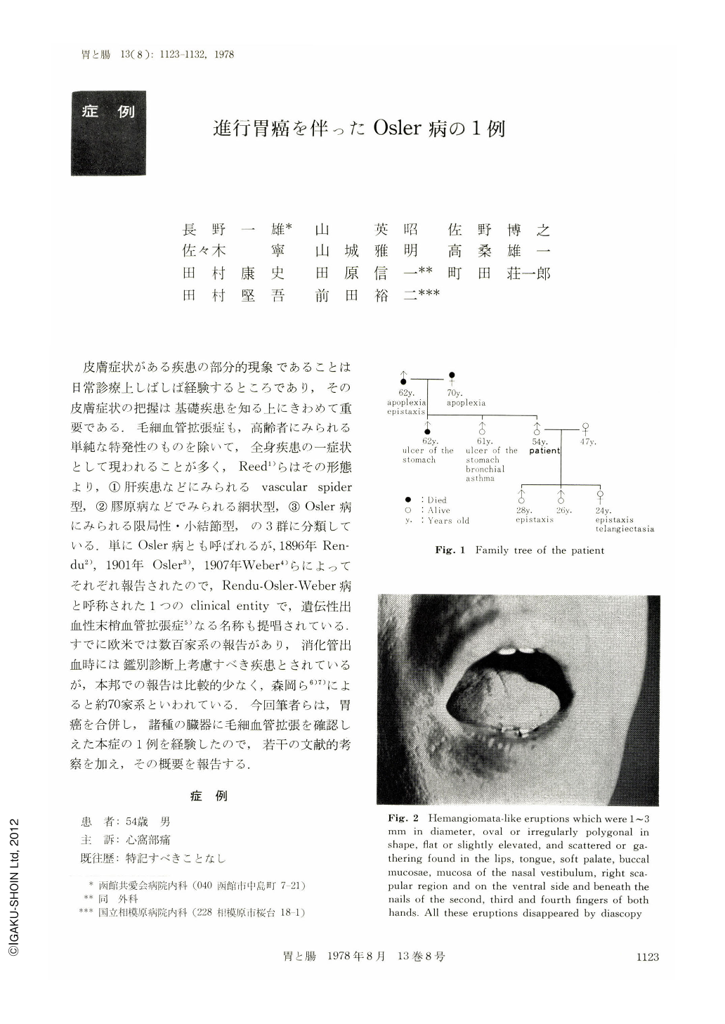
皮膚症状がある疾患の部分的現象であることは日常診療上しばしば経験するところであり,その皮膚症状の把握は基礎疾患を知る上にきわめて重要である.毛細血管拡張症も,高齢者にみられる単純な特発性のものを除いて,全身疾患の一症状として現われることが多く,Reed1)らはその形態より,①肝疾患などにみられるvascular spider型,②膠原病などでみられる網状型,③Osler病にみられる限局性・小結節型,の3群に分類している.単にOsler病とも呼ばれるが,1896年Rendu2),1901年Osler3),1907年Weber4)らによってそれぞれ報告されたので,Rendu-Osler-Weber病と呼称された1つのclinical entityで,遺伝性出血性末梢血管拡張症5)なる名称も提唱されている.すでに欧米では数百家系の報告があり,消化管出血時には鑑別診断上考慮すべき疾患とされているが,本邦での報告は比較的少なく,森岡ら6)7)によると約70家系といわれている.今回筆者らは,胃癌を合併し,諸種の臓器に毛細血管拡張を確認しえた本症の1例を経験したので,若干の文献的考察を加え,その概要を報告する.
症 例
患 者:54歳 男
主 訴:心窩部痛
既往歴:特記すべきことなし
現病歴:1952年30歳頃より鼻を強くかんだり,感冒をひいた時,あるいは鼻孔をいじったりした時などに鼻出血があり,タンポンにて容易に止血していた.この頃より口唇の恒常性の赤い点状の発疹に気づいている.1958年36歳の時,格別の誘因なく右眼視野に雲のようなものが見え,網膜硝子体出血と診断された.1975年1月および5月にも同様の右網膜硝子体出血を繰返したが,いずれも数週の加療で痕跡なく吸収された.1976年5月初旬より不定の心窩部痛が出現したが,悪心,食思不振,吐または下血,体重減少などはなかった.7月6日当科を受診し,精査のため入院した.
There are reports on hundreds of families with Osler's disease in Europe and U. S., and the disease is regarded as one which should be considered in differential diagnosis of gastrointestinal hemorrhage. On the other hand, there are relatively few reports on the families with this disease in Japan; that is, the report by Morioka et al on about 70 families is said to be the representative one. We have lately encountered one case of Osler's disease complicated by gastric cancer where telangiectasia was found in various organs. The outline of findings is presented here, together with some discussion with reference to the literature data.
A man aged 54 came to author's hospital with a chief complaint of epigastric pain. Since about 1952, he had been attacked by epistaxis after blowing the nose vigorously, or when he had caught a cold, or when he had picked the nostrils. At about that time, he had noticed the presence of red eruptions in the lips. In 1958, something like a cloud began to be visible in the right eye field. It was diagnosed as retinal bleeding. A similar retinal bleeding occurred in the right eye in January and May, 1975. On each occasion, however, the bleeding was absorbed with no trace. In July 1976, he was admitted to the hospital. A history of epistaxis was found in his father and 2 children. A history of gastric ulcer was noted in 2 of his 3 siblings. On admission, anemia was absent from the visible mucosa. Hemangioma-like eruptions were found in the lips, tongue, soft palate, buccal mucosae, mucosae of the nasal vestibulum, right scapular region and the ventral side and beneath the nails of the fingers of both hands except the little fingers. Neither any tumor nor any resistance in the abdomen was palpable. Laboratory studies disclosed that occult blood was positive in stool, but anemia was absent. The coagulative and fibrinolytic systems and hormones were all normal. No abnormalities were roentgenologically found in the digestive tract except in the stomach, where an irregularly-shaped ulcerative lesion occurred in the angular portion; the tips of the mucosal folds converging were enlarged and united. Endoscopically, hemangioma-like eruptions similar in nature to those found in the visible mucosae and skin, or dilatation of capillary vessels similar to the vascular spider were found in the mucosa of the esophagus, stomach, duodenum and rectum. At the angular portion of the stomach was observed a circular elevation with the margin shaped like a dome and with an irregularly-shaped ulceration in its center, to which the mucosal folds prominently converged. Biopsy specimen of the ulceration was diagnosed as well-differentiated adenocarcinoma. The examination of the eyegrounds disclosed angiogliosis retinae in the temporal margin of the optic disk in the right eyeground.
From these findings, this case was diagnosed as Osler's disease complicated by advanced gastric cancer. The patient underwent operation on October 9, 1976. The close observation of the abdominal cavity revealed two hemangioma-like eruptions on the surface of the right liver lobe. By macroscopic examination of the resected specimen, the ulcerative lesion was diagnosed as a Borrmann type 2 cancer and histologically as adenocarcinoma tubulare.

Copyright © 1978, Igaku-Shoin Ltd. All rights reserved.


