Japanese
English
- 有料閲覧
PPV購入案内
1,320円 (1,200円+税10%) こちらは、287ページから291ページの文献です。購入には医書.jp本会員登録が必要です。
会員登録完了後に改めて上記「購入サイトへ」を選択してください。
または「購入サイトへ」選択後に「購入ログイン」、「新規会員登録」の順に進んでください。
会員登録について詳しくはをご確認ください。
- Abstract 文献概要
- 1ページ目 Look Inside
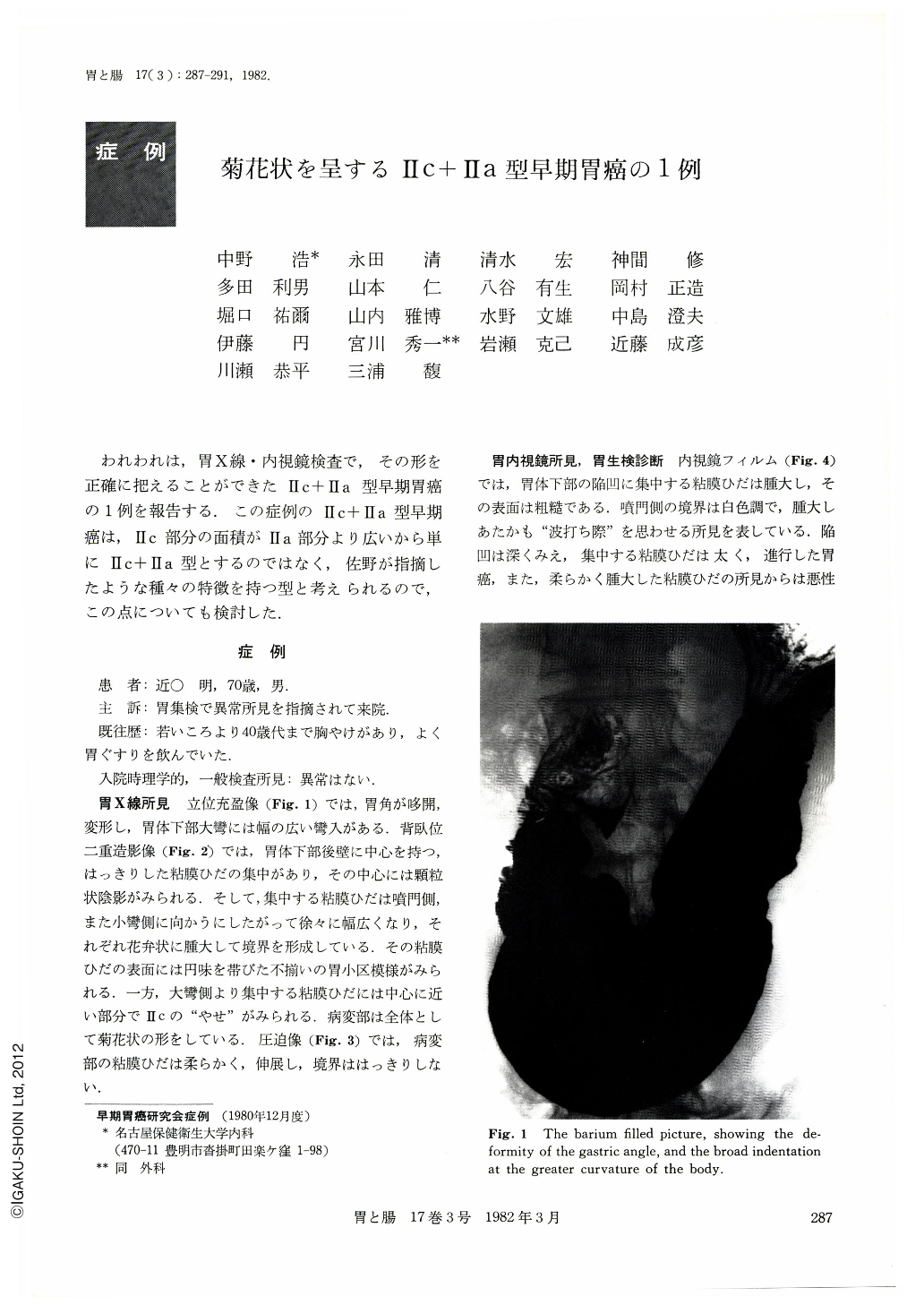
われわれは,胃X線・内視鏡検査で,その形を正確に把えることができたⅡc+Ⅱa型早期胃癌の1例を報告する.この症例のⅡc+Ⅱa型早期癌は,Ⅱc部分の面積がⅡa部分より広いから単にⅡc+Ⅱa型とするのではなく,佐野が指摘したような種々の特徴を持つ型と考えられるので,この点についても検討した.
A 70-year-old man came to our hospital for detailed examination of the stomach as he was found to have abnormal findings in the course of gastric mass survery. General and physical examinations at admission revealed no abnormality. Roentgenography of the stomach showed obvious mucosal convergency on the posterior wall of the body. In its center was seen a granular shadow. The radiating mucosal folds became gradually wider and at the margins of the lesion they looked like petals of a flower. Endoscopically the marginal part was elevated and looked white, a finding suggestive of a breaker along a beach. In the resected specimen the center of the converging folds was slightly depressed and the surrounding folds were elevated. The lesion was Ⅱc+Ⅱa type early cancer as proposed by Sano. This type of early cancer too often shows biphasic intramucosal proliferation of cancer: in the part of Ⅱa and that of Ⅱc, so that clinically it is apt to be erroneously diagnosed as advanced cancer or malignant lymphoma. In the accurate diagnosis of this type of cancer one should always bear in mind clinically possible presence of such a characteristic early cancer.
Copyright © 1982, Igaku-Shoin Ltd. All rights reserved.