Japanese
English
- 有料閲覧
- Abstract 文献概要
- 1ページ目 Look Inside
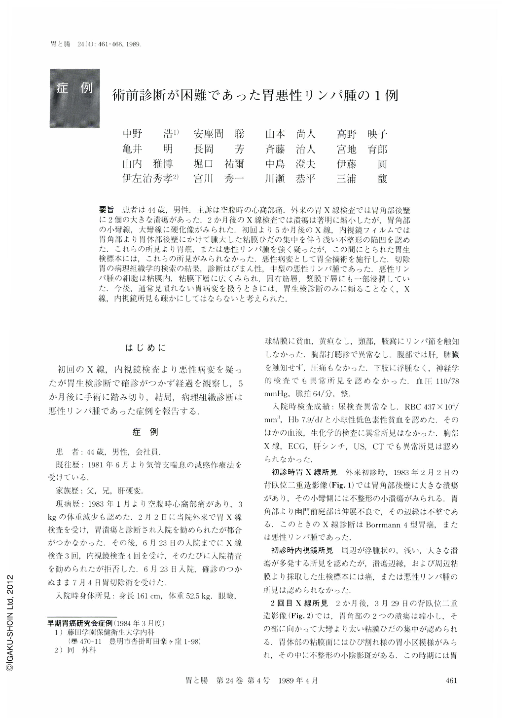
要旨 患者は44歳,男性.主訴は空腹時の心窩部痛.外来の胃X線検査では胃角部後壁に2個の大きな潰瘍があった.2か月後のX線検査では潰瘍は著明に縮小したが,胃角部の小彎線,大彎線に硬化像がみられた.初回より5か月後のX線,内視鏡フィルムでは胃角部より胃体部後壁にかけて腫大した粘膜ひだの集中を伴う浅い不整形の陥凹を認めた.これらの所見より胃癌,または悪性リンパ腫を強く疑ったが,この間にとられた胃生検標本には,これらの所見がみられなかった,悪性病変として胃全摘術を施行した.切除胃の病理組織学的検索の結果,診断はびまん性,中型の悪性リンパ腫であった.悪性リンパ腫の細胞は粘膜内,粘膜下層に広くみられ,固有筋層,漿膜下層にも一部浸潤していた.今後,通常見慣れない胃病変を扱うときには,胃生検診断のみに頼ることなく,X線,内視鏡所見も疎かにしてはならないと考えられた.
A 44-year-old man was admitted to our hospital with chief complaint of hunger epigastric pain. Physical and laboratory examinations revealed no significant abnormalities.
Routine upper GI series showed two large ulcerations in the posterior wall of the gastric angle. Two months later, these ulcerations diminished markedly in size, while the distensibility of the gastric wall decreased in the angular portion. Five months after the first examination, radiographic pictures showed shallow, large irregular-shaped depression with converging mucosal folds in the posterior wall of the gastric angle and body. Although these clinical findings highly suggested either gastric cancer or malignant lymphoma, biopsy specimen did not confirm our speculation pathologically. Total gastrectomy was carried out under the tentative diagnosis of malignant lesion.
Histological examination of the resected stomach revealed diffuse and medium-sized malignant lymphoma with extensive involvement of the mucosa and submucosa, and slight involvement of the proper muscle and subserosal layers as well. Faced with such an unusual case of gastric lesion, we should rely on not only the result of gastric biopsy but also the radiological and endoscopical features.

Copyright © 1989, Igaku-Shoin Ltd. All rights reserved.


