Japanese
English
- 有料閲覧
- Abstract 文献概要
- 1ページ目 Look Inside
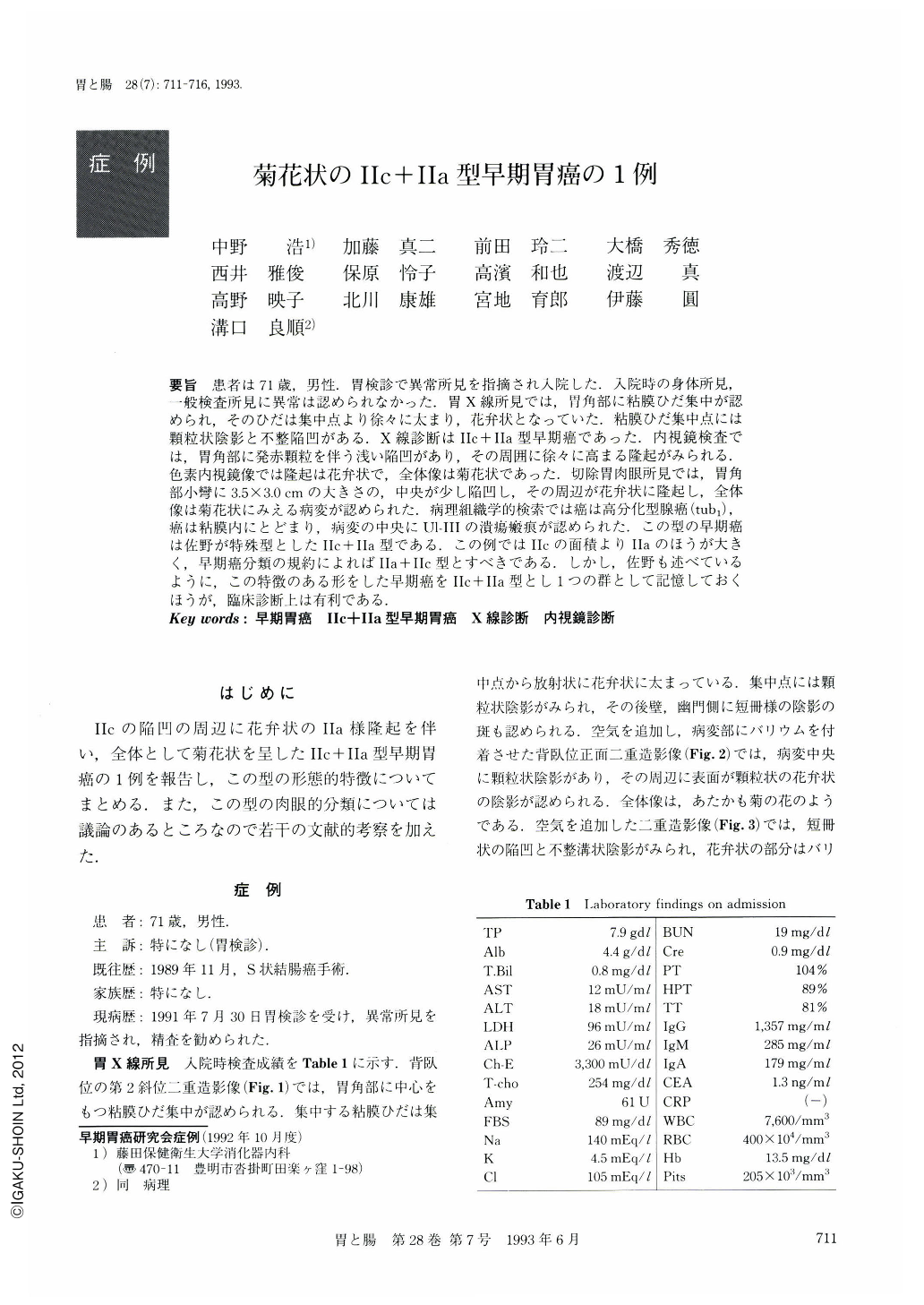
要旨 患者は71歳,男性.胃検診で異常所見を指摘され入院した.入院時の身体所見,一般検査所見に異常は認められなかった.胃X線所見では,胃角部に粘膜ひだ集中が認められ,そのひだは集中点より徐々に太まり,花弁状となっていた.粘膜ひだ集中点には顆粒状陰影と不整陥凹がある.X線診断はⅡc+Ⅱa型早期癌であった.内視鏡検査では,胃角部に発赤顆粒を伴う浅い陥凹があり,その周囲に徐々に高まる隆起がみられる.色素内視鏡像では隆起は花弁状で,全体像は菊花状であった.切除胃肉眼所見では,胃角部小彎に3.5×3.Ocmの大きさの,中央が少し陥凹し,その周辺が花弁状に隆起し,全体像は菊花状にみえる病変が認められた.病理組織学的検索では癌は高分化型腺癌(tub1),癌は粘膜内にとどまり,病変の中央にUl-Ⅲの潰瘍瘢痕が認められた.この型の早期癌は佐野が特殊型としたⅡc+Ⅱa型である.この例ではⅡcの面積よりⅡaのほうが大きく,早期癌分類の規約によればⅡa+Ⅱc型とすべきである.しかし,佐野も述べているように,この特徴のある形をした早期癌をⅡc+Ⅱa型とし1つの群として記憶しておくほうが,臨床診断上は有利である.
A 71-year-old man came to our hospital for more detailed examination, because of an abnormal finding in the screening gastric x-ray examination. Physical examination at admission showed no abnormal findings. Double contrast x-ray examination of the stomach revealed a mucosal convergence at the gastric angle. In the center of converging folds, a granular shallow depression was noted. The radiating folds became gradually wider with their distance from the cencer and looked like flower petals. Radiological diagnosis was a type Ⅱc+Ⅱa early gastric cancer.
Endoscopically, the lesion mimicked a flower of the chrysanthemum. In the resected specimen, a type Ⅱc+Ⅱa lesion, 3.5×3.0 cm in size, was located on the lesser curvature at the gastric angle. Histological diagnosis was well differentiated adenocarcinoma with an ulcer scar in the center of the lesion. Cancer invasion was limited within the mucosa. We classified this lesion as a type Ⅱc+Ⅱa early gastric cancer according to the original classification of Dr. Sano. According to the rule for macroscopic classification of early cancer, this lession should be classified into type Ⅱa+Ⅱc, because an area of type Ⅱa was bigger than that of type Ⅱc. We dare to use type Ⅱc+Ⅱa. Because of its characteristic shape, it would be useful to identify this type of lesions as an independent group.

Copyright © 1993, Igaku-Shoin Ltd. All rights reserved.


