Japanese
English
- 有料閲覧
PPV購入案内
1,320円 (1,200円+税10%) こちらは、245ページから251ページの文献です。購入には医書.jp本会員登録が必要です。
会員登録完了後に改めて上記「購入サイトへ」を選択してください。
または「購入サイトへ」選択後に「購入ログイン」、「新規会員登録」の順に進んでください。
会員登録について詳しくはをご確認ください。
- Abstract 文献概要
- 1ページ目 Look Inside
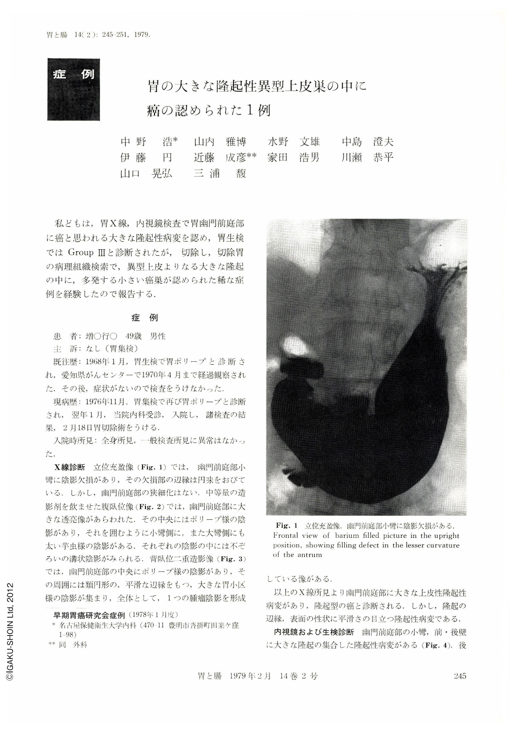
私どもは,胃X線,内視鏡検査で胃幽門前庭部に癌と思われる大きな隆起性病変を認め,胃生検ではGroup Ⅲと診断されたが,切除し,切除胃の病理組織検索で,異型上皮よりなる大きな隆起の中に,多発する小さい癌巣が認められた稀な症例を経験したので報告する.
Radiological and endoscopical examinations demonstrated a large cancerous protruded lesion in the antrum of the stomach. The margin of the elevation was, however, smooth radiologically, and white colored, discoid protrusion characteristic of the so-called atypical epithelium was seen endoscopically in a part of the large elevation. The findings made it difficult to diagnose the whole lesion as cancer.
The biopsy specimen presented histologic feature of Group Ⅲ which indicated borderline malignancy. The resected stomach showed a regularly shaped protrusion with soft nodular surface, measuring 5 cm×7 cm.
The nodular elevations were either flower-bed like or polypoid. The macroscopic findings highly suggested malignancy. Histological examination of the resected stomach revealed that the elevation consisted of adenomatous changes with atypical tubular structure and in part, papillary hyperplasia. Cancer was detected in a limited narrow area.
The present case is interesting from the standpoint of the occurrence of cancer in an exceptionally large lesion of atypical epithelia or the probable cancerous change of adenomatous polyp.
Copyright © 1979, Igaku-Shoin Ltd. All rights reserved.