Japanese
English
- 有料閲覧
- Abstract 文献概要
- 1ページ目 Look Inside
- サイト内被引用 Cited by
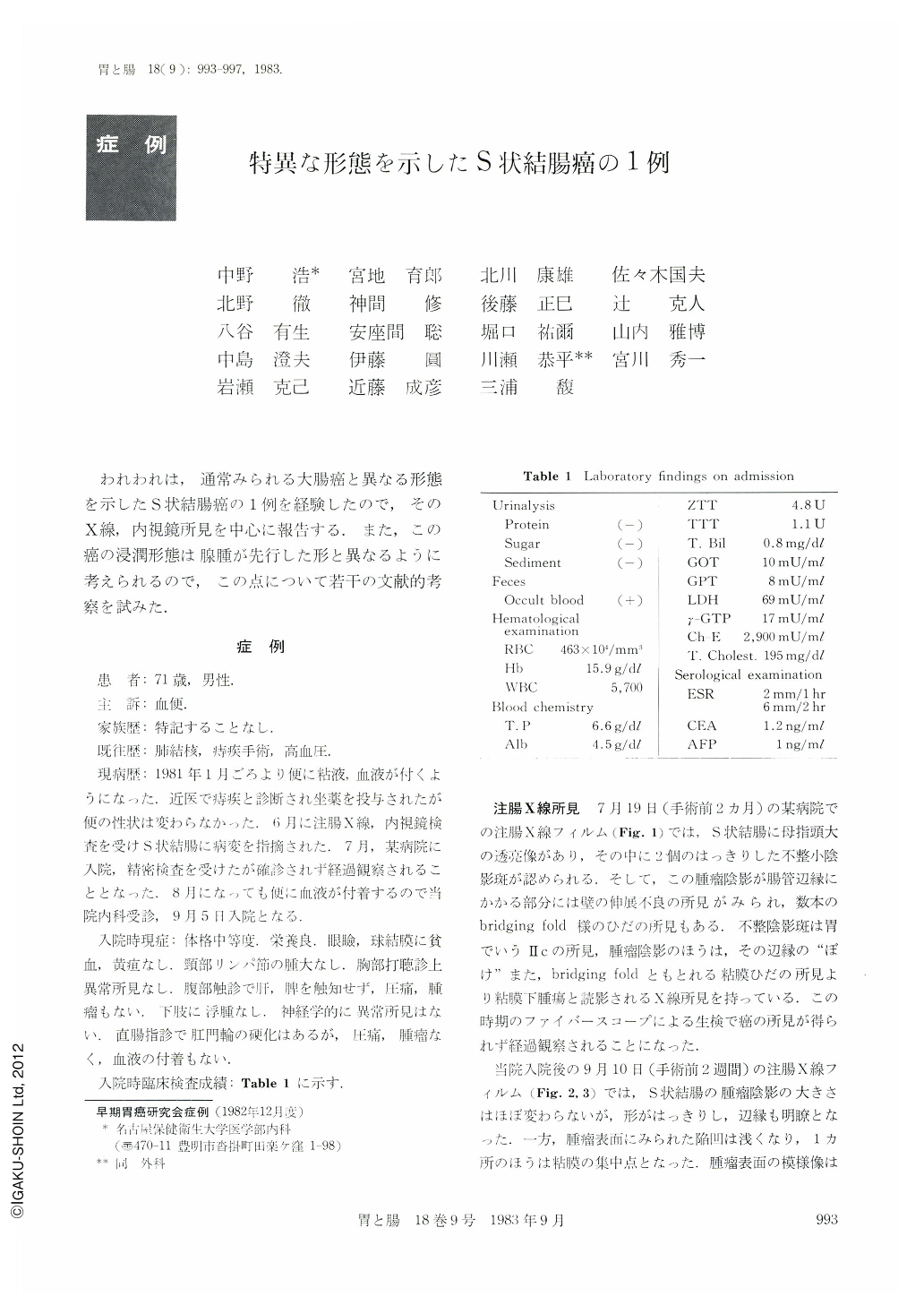
われわれは,通常みられる大腸癌と異なる形態を示したS状結腸癌の1例を経験したので,そのX線,内視鏡所見を中心に報告する.また,この癌の浸潤形態は腺腫が先行した形と異なるように考えられるので,この点について若干の文献的考察を試みた.
症 例
患 者:71歳,男性.
主 訴:血便.
家族歴:特記することなし.
既往歴:肺結核,痔疾手術,高血圧.
現病歴:1981年1月ごろより便に粘液,血液が付くようになった,近医で痔疾と診断され坐薬を投与されたが便の性状は変わらなかった.6月に注腸X線,内視鏡検査を受けS状結腸に病変を指摘された.7月,某病院に入院,精密検査を受けたが確診されず経過観察されることとなった.8月になっても便に血液が付着するので当院内科受診,9月5日入院となる.
A 71 year-old man was admitted to our hospital with a chief complaint of bloody stool.
Barium enema examination revealed a small polypoid lesion with irregular-shaped depression in the sigmoid colon.
Colonofiberscopy visualized a lobulated polypoid lesion with reddish central depression. The surface of the lesion was the same mucosa as that adjacent to the lesion. Although the biopsy specimen showed adenocarcinoma, the polypoid lesion apparently differed from the typical radiologic and endoscopic findings of colonic cancer.
The resected specimen showed a discoid mass, measurfing 2.0 cm×1.8 cm in size, with multiple small depressions on the surface which was covered with the same mucosa as that adjacent to the lesion.
In the section of the resected material, cancer was located in the erosive and ulcerated area of the mucosal layer, and cancerous invasion as well as proliferation formed a tumor mass in the submucosal layer. A part of the cancer tissue extended through the proper muscle layer to the subserosal layer.
There was no evidence of adenoma in this colonic cancer tissue, and the findings of the cross section differed largely from those of usual colon cancer which is believed to occur in association with preceding adenoma.

Copyright © 1983, Igaku-Shoin Ltd. All rights reserved.


