Japanese
English
- 有料閲覧
PPV購入案内
1,320円 (1,200円+税10%) こちらは、1247ページから1252ページの文献です。購入には医書.jp本会員登録が必要です。
会員登録完了後に改めて上記「購入サイトへ」を選択してください。
または「購入サイトへ」選択後に「購入ログイン」、「新規会員登録」の順に進んでください。
会員登録について詳しくはをご確認ください。
- Abstract 文献概要
- 1ページ目 Look Inside
筆者らは最近67歳の女性にみられた右側回結腸炎の手術症例を経験したので報告し,若干の考察をこころみた.
患者は初発症状の発現後一旦寛解していたが,約2年後に再燃増悪を来したため,罹患部腸管の切除術が施行された.術後の病理組織学的検査では,非特異性肉芽腫性炎症性病変であることが明らかにされた.
A 67-year-old female was admitted with an attack of recurrent bloody diarrhea. About two years before admission, she had the first attack of melena. Surgical treatment was recommended after clinical examinations which disclosed the exacervation of so-called right sided ileocolitis. A right hemicolectomy and ileotransverse colostomy were performed. The postoperative histological studies of the resected speciemen revealed the lesion composed of non-specific granulomatous infiammation with multiple irregular colonic (or ileal) ulcers.
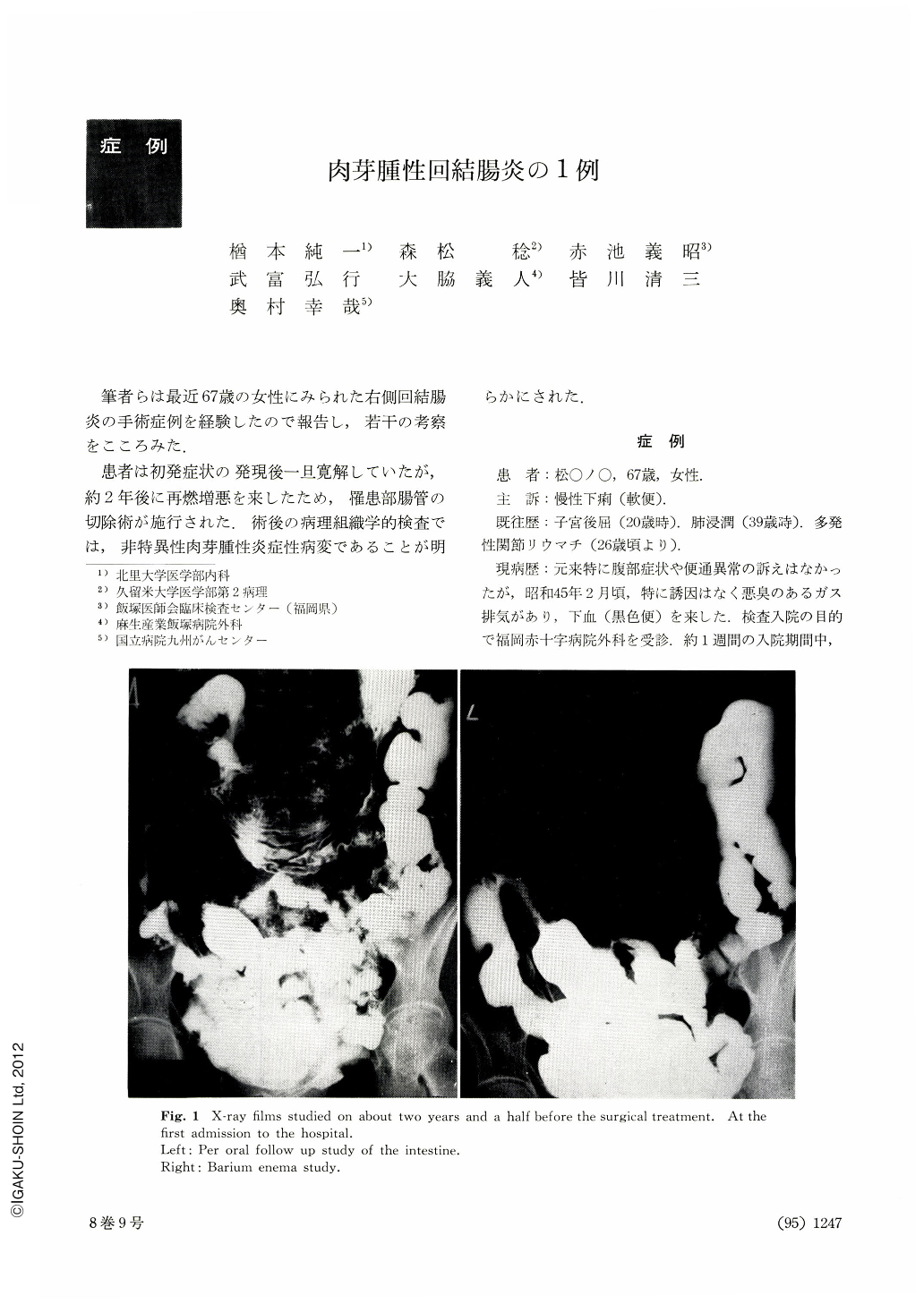
X-ray examinations: Barium enema revealed that the cecum, ascending colon, proximal part of the transverse colon, and terminal ileum were involved. Multiple irregular ragged ulcers and pseudopolyposis-like cobble stone appearances were seen. Involved bowel wall was not so thickened nor indurated. Spastic movement of the involved bowel was observed. Narrowing was seen in the distal part of the involved colon. Ileal involvement with dilated terminal ileum was so short as about five centimeters. Skip lesions were not observed ileum, were normal.
An upper gastrointestinal series and small bowel follow-up, excluding the terminal ileum, were normal.
Chest x-ray film showed no remarkable findings.
Pathological findings: Gross appearance was corre-sponded with barium enema findings. Microscopically, the ragged ulcers were composed of Ul-II undermining ulcers with marked proliferation of lymphocytic aggregates. Submucosal fibrosis, lymphocytic aggregates and non-caseating tuberculoid granulomas were charactaristics of the lesion. Stains for mycobacterium tuberculosis were negative. Lymphocytic aggregates and tuberculoid granulomas were occasionally observed in all coats of the involved area.
Copyright © 1973, Igaku-Shoin Ltd. All rights reserved.