Japanese
English
- 有料閲覧
- Abstract 文献概要
- 1ページ目 Look Inside
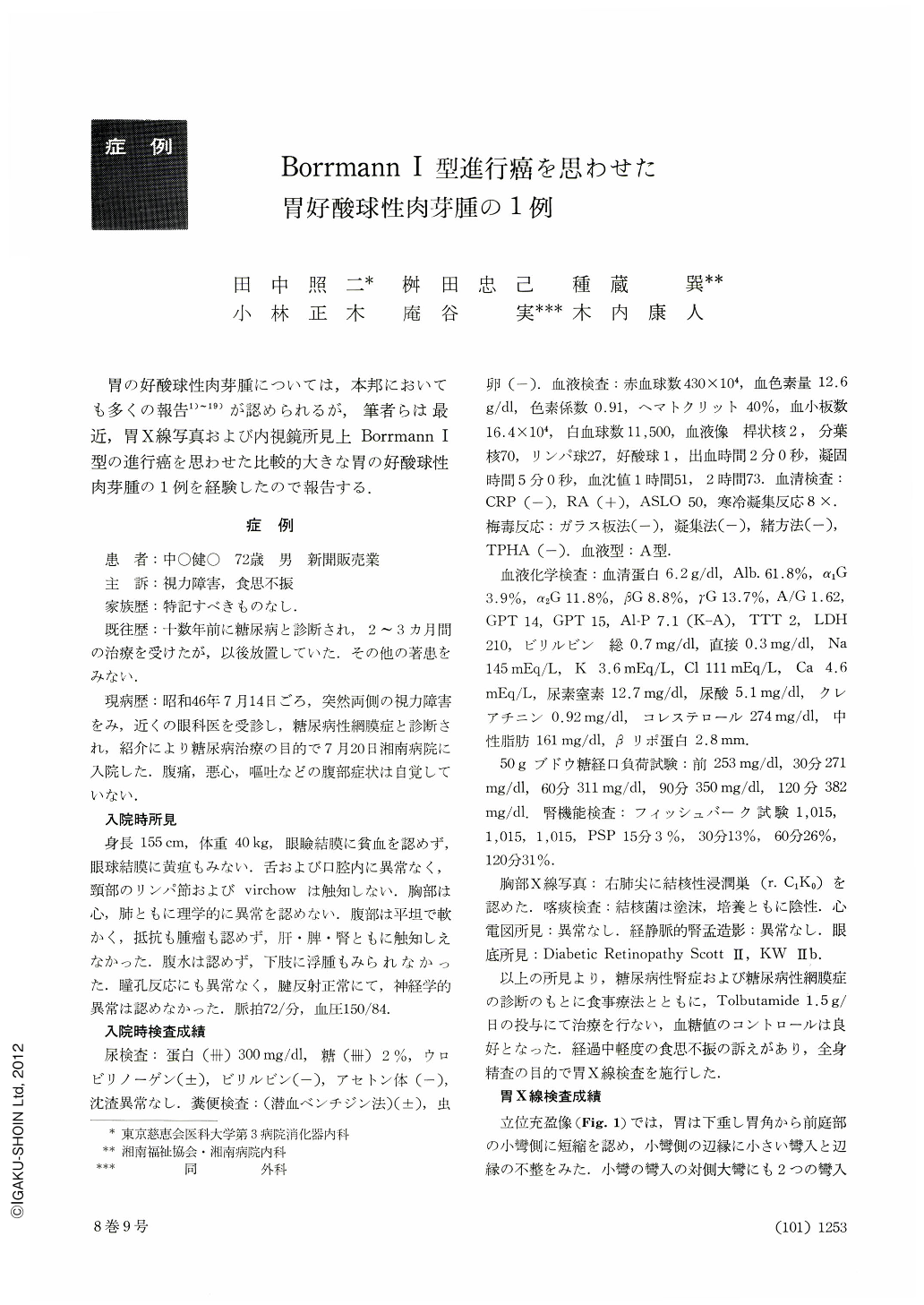
胃の好酸球性肉芽腫については,本邦においても多くの報告1)~19)が認められるが,筆者らは最近,胃X線写真および内視鏡所見上Borrmann Ⅰ型の進行癌を思わせた比較的大きな胃の好酸球性肉芽腫の1例を経験したので報告する.
A 72 years old male was admitted to the Shonan Hospital on July 14, 1971 to be treated diabetes mellitus by suggestion of a ophthalmologist. During the treatment with tolbutamide, a G-I series was performed as he complained anorexia. And as a consequence of G-I series, a chestnut sized oval shadow defect was found at the antrum of stomach with irregular contour of the lesser curvature in a upright, barium-filled picture. Photographs by gastrocamera also showed a fairly large sized polypoid with white fur of uneven surface.
Under the diagnosis of advanced gastric cancer of Borrmann I Type, laparotomy was performed. As surrounding mucosal finding of the stalk was normal, fairly large tumor (3×6 cm) with the stalk was extirpated.
Histologically, the tumor consisted mainly of hyperplasia of the connective tissues and diffuse infiltration of eosinophilic cells, accompanied with formation of lymph follicles. Any parasite was not found and then this case can be diagnosed as an inflammatory fibroid polyp, namely “localized eosinophilic granuloma”.

Copyright © 1973, Igaku-Shoin Ltd. All rights reserved.


