Japanese
English
- 有料閲覧
- Abstract 文献概要
- 1ページ目 Look Inside
著者らが最近4年半Dec. 1, 1966.-May. 31, 1971.に取り扱った切除胃の病理組織学的検査症例は254例であるが,そのうち胃好酸球性肉芽腫が4例(1.6%)含まれていた.3例は寄生虫によらないもので,1例は寄生虫(アニサキス)によるものであった.自験例について比較検討し,若干の知見を得たので報告する.
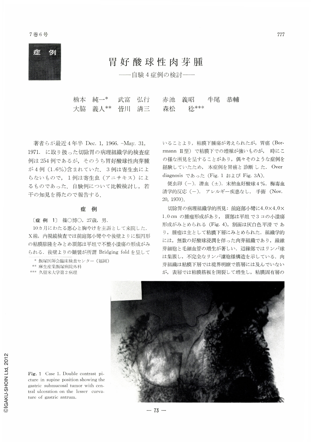
Case 1: A 27-years-old male had been suffering from stomach trouble for about 10 months. Upper gastrointestinal series disclosed a gastric tumor with central ulceration on the lesser curvature of the gastric antrum. Histologically, the tumor was composed of eosinophilic glanuloma measuring 4.0 by 4.0 by 1.0 cm.
Case 2: A 54-years-old male was admitted, complainning of intractable epigastralgia. Upper gastrointestinal series disclosed a large ulcerative lesion associated with submucosal tumor. Histologically, eosinophilic granuloma was found diffusely in both the ulcer edge and bottom. The granuloma measured 6.0 by 4.0 by 1.0 cm, and the ulcer, 4.5 by 3.5 by 0.5 cm.
Case 3: A 70-years-old female was admitted with a recurrent attack of cholelithiasis, and was recommended cholecystectomy. Preoperative routine examinations of the upper gastrointestinal tract disclosed a gastric polyp on the posterior wall of the antrum. Histologically, the polyp, measuring 1.5 by 1.2 by 1.2 cm, was composed of eosinophilic granuloma (inflammatory fibroid polyp).
Case 4: A 47-years-old male underwent gastrectomy because of uncurable peptic ulcers. Two gastric submucosal tumors were incidentally detected on the resected stomach. Histologically, both tumors were composed of parasitic granulomas with eosinophilic infiltration.
There were neither parasitic bodies nor localized abscesses in cases 1, 2 and 3. On the other hand, granulomas were situated mainly in the submucosa but extended into the muscular layers in case 2 and 4. Muscularis mucosae was split into isolated bundles in case 1 and 3. On the other hand, mucularis mucosae was maintained in the both tumors of case 4. Case 2 was thought to be a rare type of gastric eosinophilic granuloma.

Copyright © 1972, Igaku-Shoin Ltd. All rights reserved.


