Japanese
English
- 有料閲覧
PPV購入案内
1,320円 (1,200円+税10%) こちらは、931ページから935ページの文献です。購入には医書.jp本会員登録が必要です。
会員登録完了後に改めて上記「購入サイトへ」を選択してください。
または「購入サイトへ」選択後に「購入ログイン」、「新規会員登録」の順に進んでください。
会員登録について詳しくはをご確認ください。
- Abstract 文献概要
- 1ページ目 Look Inside
胃の良性lymphoid lesionは一般に胃RLH(reactive lymphoreticular hyperplasia)と呼ばれ,臨床的には胃癌との鑑別診断において,また病理組織学的には胃悪性リンパ腫との関連性において興味ある病変である.
著者らは最近胃粘膜下腫瘍状を呈し,病理組織学的にも特異な所見をみとめたいわゆる胃RLHの2症例を経験したので報告する.
The purpose of this paper is to report two cases of localized reactive Lymphoreticular hyperplasia of the stomach and compare them to the previous literature.
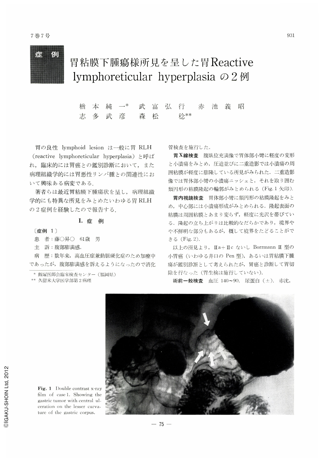
Case 1: A 61 years old male had complaint of epigastric fullness, while treated for arteriosclerosis with hypertension. Upper gastrointestinal series disclosed a gastric tumor with central ulceration on the lesser curvature of the gastric corpus. Clinically, it was difficult to clarify its nature, benign or malignant. Histologically, the tumor, measuring 3.0 by 2.0 by 0.8 cm, was composed of massive lymphoid hyperplasia situated mainly in the submucosa infiltrating down to the muscular layer with relative clear-cut border. Lymphoid hyperplasia with reaction centers was found in the lesion. Infiltrating cells were composed of lymphocytes, reticulum cells and inflammatory cells without cytological malignancies. No remarkable changes were seen in the lymph nodes. There were no evidences of malignancy during fifteenth months' follow-up period.
Case 2: A 56 years old female was admitted with an attack of eholelithiasis. Surgical treatment was recommended after clinical examinations such as liver function test, cholecystography and upper gastrointestinal series. X-ray of the stomach disclosed a gastric submucosal tumor on the anterior wall of the lower cardiac area. Cholecytectomy combined with gastrectomy was performed. Postoperative histological study of the stomach revealed the tumor, measuring 3.0 by 2.5 by 1.0 cm, composed of Lymphoreticular hyperplasia with proliferation of collagen fibers.
There is no single criterion which helps to make accurate the differential diagnosis of malignant lymphoma and massive lymhoid hyperplasia such as in case 1. The final diagnosis may depend upon the followup study after operation.
Copyright © 1972, Igaku-Shoin Ltd. All rights reserved.