Japanese
English
- 有料閲覧
PPV購入案内
1,320円 (1,200円+税10%) こちらは、971ページから975ページの文献です。購入には医書.jp本会員登録が必要です。
会員登録完了後に改めて上記「購入サイトへ」を選択してください。
または「購入サイトへ」選択後に「購入ログイン」、「新規会員登録」の順に進んでください。
会員登録について詳しくはをご確認ください。
- Abstract 文献概要
- 1ページ目 Look Inside
- サイト内被引用 Cited by
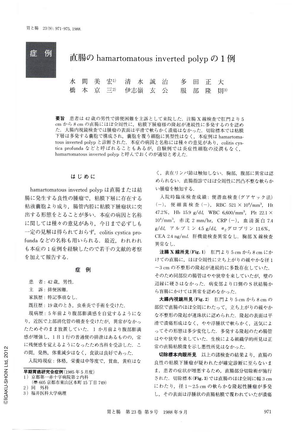
要旨 患者は42歳の男性で排便困難を主訴として来院した。注腸X線検査で肛門より5cmから8cmの直腸にほぼ全周性に,粘膜下腫瘤様の隆起が連続性に多発するのを認めた.大腸内視鏡検査では腫瘤の表面は平滑で軟らかく潰瘍はなかった.切除標本では粘膜下層は多発する囊胞で構成され,囊胞を覆う細胞に異型性はなく,本症例はhamartomatous inverted polypと診断された.本症の病因と名称には種々の意見があり,colitis cystica profundaなどと呼ばれることもあるが,自験例では炎症性細胞の浸潤もなく,hamartomatous inverted polypと呼んでおくのが適切と考えた.
A 42-year-old man came to our hospital complaining of constipation. Digital examination of the rectum disclosed a narrowing at 5 cm from the anal sphincter. Laboratory data were nonspecific. Barium enema revealed a narrow segment with nodularity (Fig. 1). Colonoscopy disclosed an aggregate of nodular protrusions extending from 5 to 8 cm from the anal sphincter (Fig. 2). The lesion was covered with normal mucosa with the biopsy specimen demonstrating no malignant cells. Subsequently, partial resection of the rectum was carried out (Fig. 3). Histologically the lesion consists of mucin-containing cysts in the submucosa (Fig. 4). It was lined by tall columnar epithelium and surrounded by muscularis mucosa (Fig. 5). Because of the lack of inflammatory changes, the lesion was considered as hamartomatous inverted polyp rather than colitis cystica profunda.
Copyright © 1988, Igaku-Shoin Ltd. All rights reserved.