Japanese
English
- 有料閲覧
PPV購入案内
1,320円 (1,200円+税10%) こちらは、976ページから980ページの文献です。購入には医書.jp本会員登録が必要です。
会員登録完了後に改めて上記「購入サイトへ」を選択してください。
または「購入サイトへ」選択後に「購入ログイン」、「新規会員登録」の順に進んでください。
会員登録について詳しくはをご確認ください。
- Abstract 文献概要
- 1ページ目 Look Inside
- サイト内被引用 Cited by
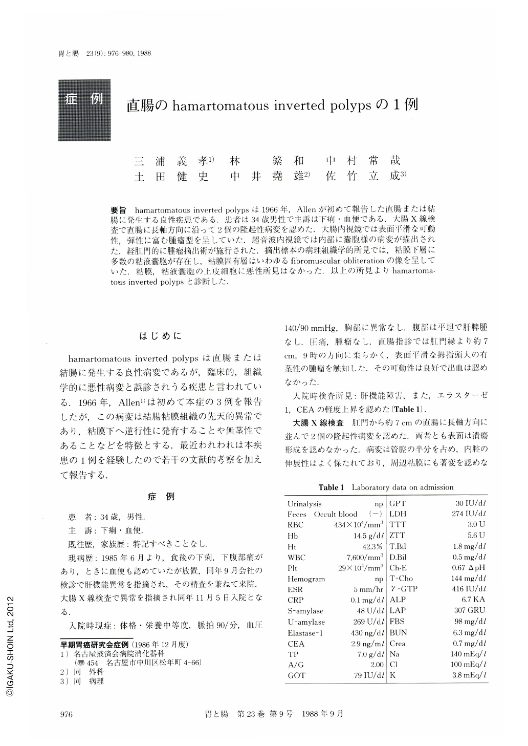
要旨 hamartomatous inverted polypsは1966年,Allenが初めて報告した直腸または結腸に発生する良性疾患である.患者は34歳男性で主訴は下痢・血便である.大腸X線検査で直腸に長軸方向に沿って2個の隆起性病変を認めた.大腸内視鏡では表面平滑な可動性,弾性に富む腫瘤型を呈していた.超音波内視鏡では内部に囊胞様の病変が描出された,経肛門的に腫瘤摘出術が施行された.摘出標本の病理組織学的所見では,粘膜下層に多数の粘液囊胞が存在し,粘膜固有層はいわゆるfibrornuscular obliterationの像を呈していた.粘膜,粘液囊胞の上皮細胞に悪性所見はなかった.以上の所見よりhamartomatous inverted polypsと診断した.
Hamartomatous inverted polyp is a rare disease that was first described by Allen in 1966.
A 34 year-old man was admitted to Nagoya Ekisaikai Hospital on November 5, 1985, for complaints of diarrhea and rectal bleeding.
On barium enema two elevated lesions were found in the rectum (Fig. 1). Endoscopy confirmed these two lesions located along the longitudinal axis of the rectum and covered with normal appearing mucosa (Fig. 2, 3).
Endoscopic ultrasonography revealed many cysts in the submucosa (Fig. 4).
Local excision was performed. Microscopically, many cysts covered with columnar epithelium without malignant transformation were seen in the submucosa. The lamina propria was occupied by excessive amount of collagen fibers and smooth muscle cells showing fibromuscular obliteration (Fig. 6a, b). Pathologic study revealed colitis cystica profunda.
Young age, no previous history of inflammatory bowel disease, and the lack of inflammatory reaction around the lesions as well as the histological findings as above formed the basis of diagnosing this case as hamartomatous inverted polyps.
Copyright © 1988, Igaku-Shoin Ltd. All rights reserved.