Japanese
English
- 有料閲覧
PPV購入案内
1,320円 (1,200円+税10%) こちらは、1259ページから1265ページの文献です。購入には医書.jp本会員登録が必要です。
会員登録完了後に改めて上記「購入サイトへ」を選択してください。
または「購入サイトへ」選択後に「購入ログイン」、「新規会員登録」の順に進んでください。
会員登録について詳しくはをご確認ください。
- Abstract 文献概要
- 1ページ目 Look Inside
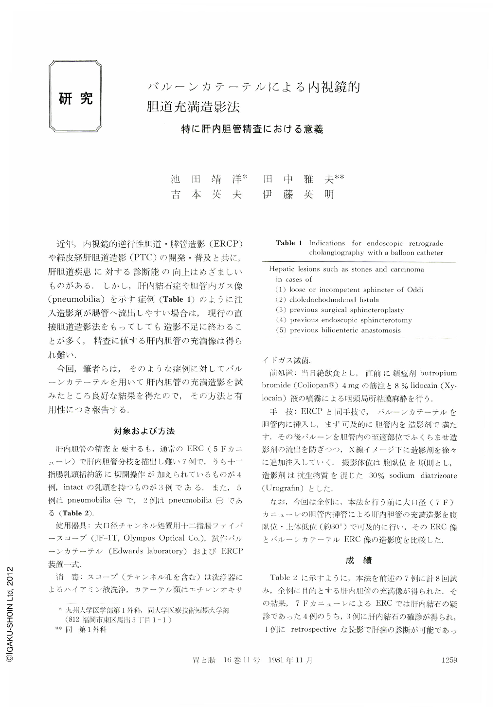
近年,内視鏡的逆行性胆道・膵管造影(ERCP)や経皮経肝胆道造影(PTC)の開発・普及と共に,肝胆道疾患に対する診断能の向上はめざましいものがある.しかし,肝内結石症や胆管内ガス像(pneumobilia)を示す症例(Table 1)のように注入造影剤が腸管へ流出しやすい場合は,現行の直接胆道造影法をもってしても造影不足に終わることが多く,精査に値する肝内胆管の充満像は得られ難い.
今回,筆者らは,そのような症例に対してバルーンカテーテルを用いて肝内胆管の充満造影を試みたところ良好な結果を得たので,その方法と有用性につき報告する.
In seven patients whose hepatic ducts were not visualized by standard endoscopic cholangiography with a 5 French catheter, hepatic duct filling was attempted by injection of contrast medium using a large-calibre catheter of 7 French size and an end. hole balloon catheter passed through the Olympus JF-1T duodenoscope. The reasons for the poor visualization of the hepatic ducts were an overflow of injected contrast medium through the patulous sphincter in two patients, the endoscopic sphincterotomy orifice in three, the sphincteroplasty in one and the cholecystoduodenostomy in one. Cholangiography with the 7F catheter showed indefinite findings suggesting the possible presence of hepatic duct stones in four patients, but stones were clearly visualized by cholangiography with the balloon catheter in three of these patients. In another patient, the obstruction of the left hepatic duct associated with rigidity and irregularity of fthe bile duct wall was demonstrated by balloon cholangiography. It proved to be due to carcinoma by subsequent laparotomy. The hepatic branches were also well visualized in the remaining three patients and the absence of hepatic duct stones was confirmed.
Copyright © 1981, Igaku-Shoin Ltd. All rights reserved.