Japanese
English
- 有料閲覧
PPV購入案内
1,320円 (1,200円+税10%) こちらは、1127ページから1132ページの文献です。購入には医書.jp本会員登録が必要です。
会員登録完了後に改めて上記「購入サイトへ」を選択してください。
または「購入サイトへ」選択後に「購入ログイン」、「新規会員登録」の順に進んでください。
会員登録について詳しくはをご確認ください。
- Abstract 文献概要
- 1ページ目 Look Inside
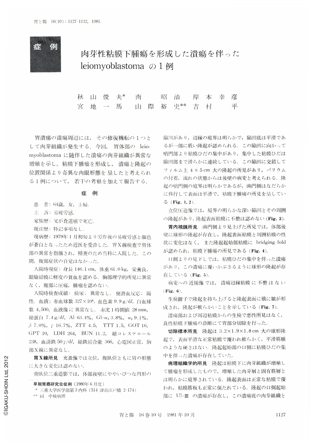
胃潰瘍の潰瘍周辺には,その修復機転の1つとして肉芽組織が発生する.今回,胃体部のleiomyoblastomaに随伴した潰瘍の肉芽組織が異常な増殖を示し,粘膜下腫瘤を形成し,潰瘍と隆起の位置関係より奇異な肉眼形態を呈したと考えられる1例について,若干の考察を加えて報告する.
A64-year-old woman underwent gastric examination because of iron deficiency anemia. A submucosal tumor 5×4 cm in diameters was seen on the posterior wall of the body. The endoscopic finding was peculiar because a deep ulcer was seen as if the oral base of the protrusion had been scooped out. The ulcer margings and converging folds were not irregular. As biopsy was also negative for malignancy, partial gastrectomy was performed under a diagnosis of a benign tumor.
Histologically, the submucosal protrusion was caused by proliferation of granulomatous tissue, continuous with that of the ulcer base. Extraordinary proliferation of granulomatous tissue in the process of ulcer healing must have produced such asubmucosal tumor. The granulomatous tissue was edematous and of soft consistency without any eosinophilic infiltration. Partially the muscularis propria in the region of ulcer was deeply stared by H・E. It was also displaced partly by the granulomatous tissue. Here was recognized a small leiomyoblastoma. Ulcer was considered to have originated by this tumor.
Ulcer on the submucosal tumor often occurs on the top of the protrusion, but in this case the peculiar macroscopic finding was caused by the presence of ulcer in the beginning of the protrusion as reactive granulomatous tissue due to ulcer had formed a submucosal protrusion.
We believe that extraordinary proliferation of granulation is related with the occurrence of ulcer in the relatively sparse submucosal tissue in the side of the greater curvature and also with the patient's constitution.
Copyright © 1981, Igaku-Shoin Ltd. All rights reserved.