Japanese
English
- 有料閲覧
PPV購入案内
1,320円 (1,200円+税10%) こちらは、437ページから440ページの文献です。購入には医書.jp本会員登録が必要です。
会員登録完了後に改めて上記「購入サイトへ」を選択してください。
または「購入サイトへ」選択後に「購入ログイン」、「新規会員登録」の順に進んでください。
会員登録について詳しくはをご確認ください。
- Abstract 文献概要
- 1ページ目 Look Inside
近年胃のX線および内視鏡診断技術の進歩に伴い,胃粘膜下腫瘍の発見ならびに質的診断も向上し,その報告例が数多く見られるようになった.bizarre leiomyoblastomaも術後の病理学的検索によって発見された症例は幾らか報告が見られるが,術前に質的確定診断のなされた報告はまだ見られない.われわれは最近本症を経験したので報告する.
A 44-year-old male began to have hunger epigastralgia in May 1973 and was hospitalized 1 week later for minute examination.
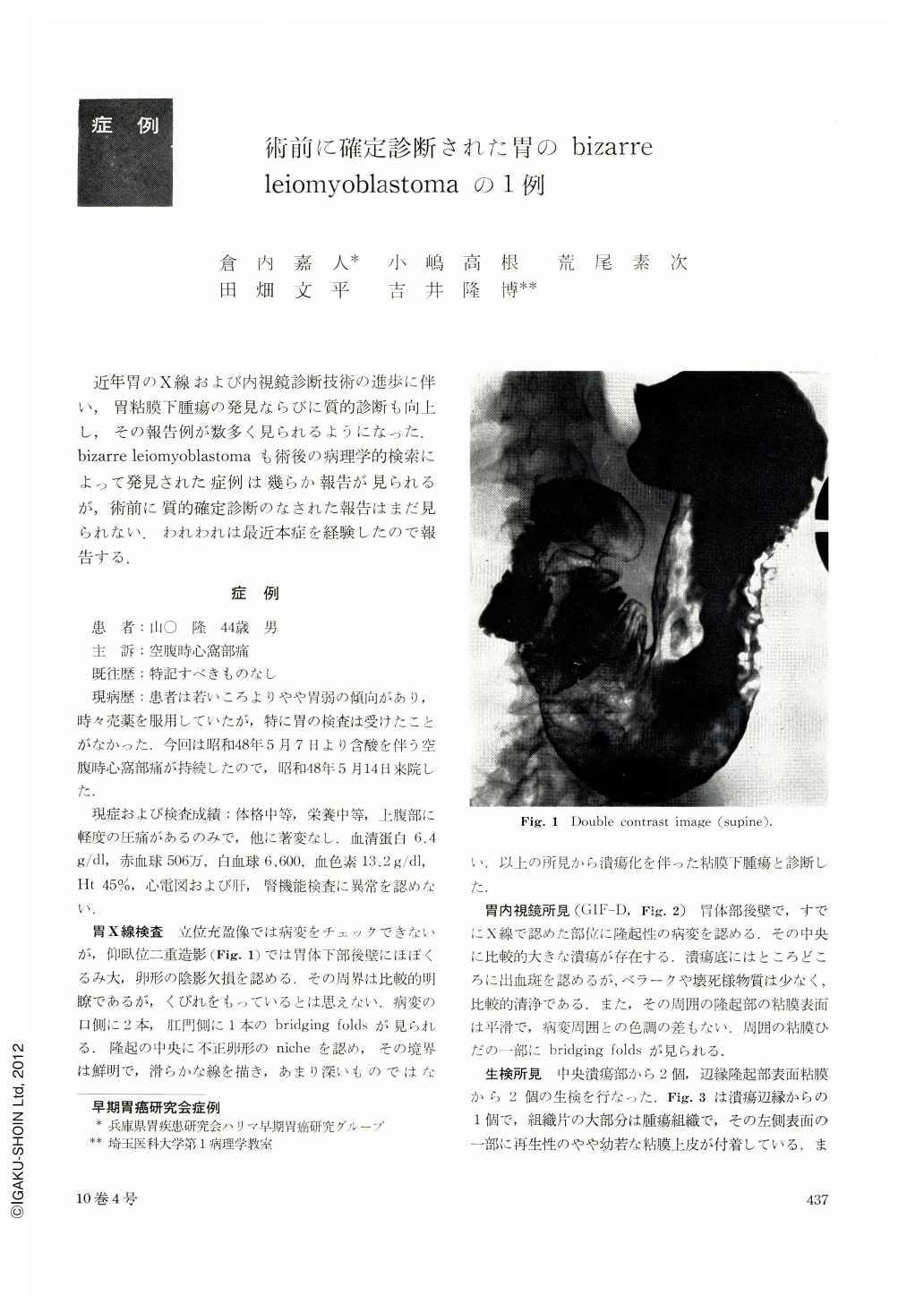
Double contrast image in supine position revealed a well-defined elevated lesion with a central niche in the posterior wall of gastric body. A few bridging folds were noted. With gastrofiberscopy, the lesion was found to be covered by the smooth mucosa having usual color. The base of ulcer demonstrated some hemorrhagic foci and a relatively small amount of necrotic material covering. Two biopsy materials obtained from the margin of ulcer showed neoplastic tissue. The tumor cells were arranged densely and solidly without alveolar structure. The majority of tumor cells were spherically swollen by vacuolar degeneration. Special stainings for glycogen, mucus and fat revealed negative results. Some tumor cells were spindle in shape, representing smooth-muscle character. From these findings, the lesion was diagnosed as bizarre leiomyoblastoma. From Stout's criteria, the lesion was regarded to be benign, because mitosis was scarcely seen.
Gastrectomy was carried out on May 31, 1973. The tumor was located mainly in the submucosa and partly in the proper muscle coat and considered to have arisen from the proper muscle coat.
Copyright © 1975, Igaku-Shoin Ltd. All rights reserved.