Japanese
English
- 有料閲覧
PPV購入案内
1,320円 (1,200円+税10%) こちらは、657ページから662ページの文献です。購入には医書.jp本会員登録が必要です。
会員登録完了後に改めて上記「購入サイトへ」を選択してください。
または「購入サイトへ」選択後に「購入ログイン」、「新規会員登録」の順に進んでください。
会員登録について詳しくはをご確認ください。
- Abstract 文献概要
- 1ページ目 Look Inside
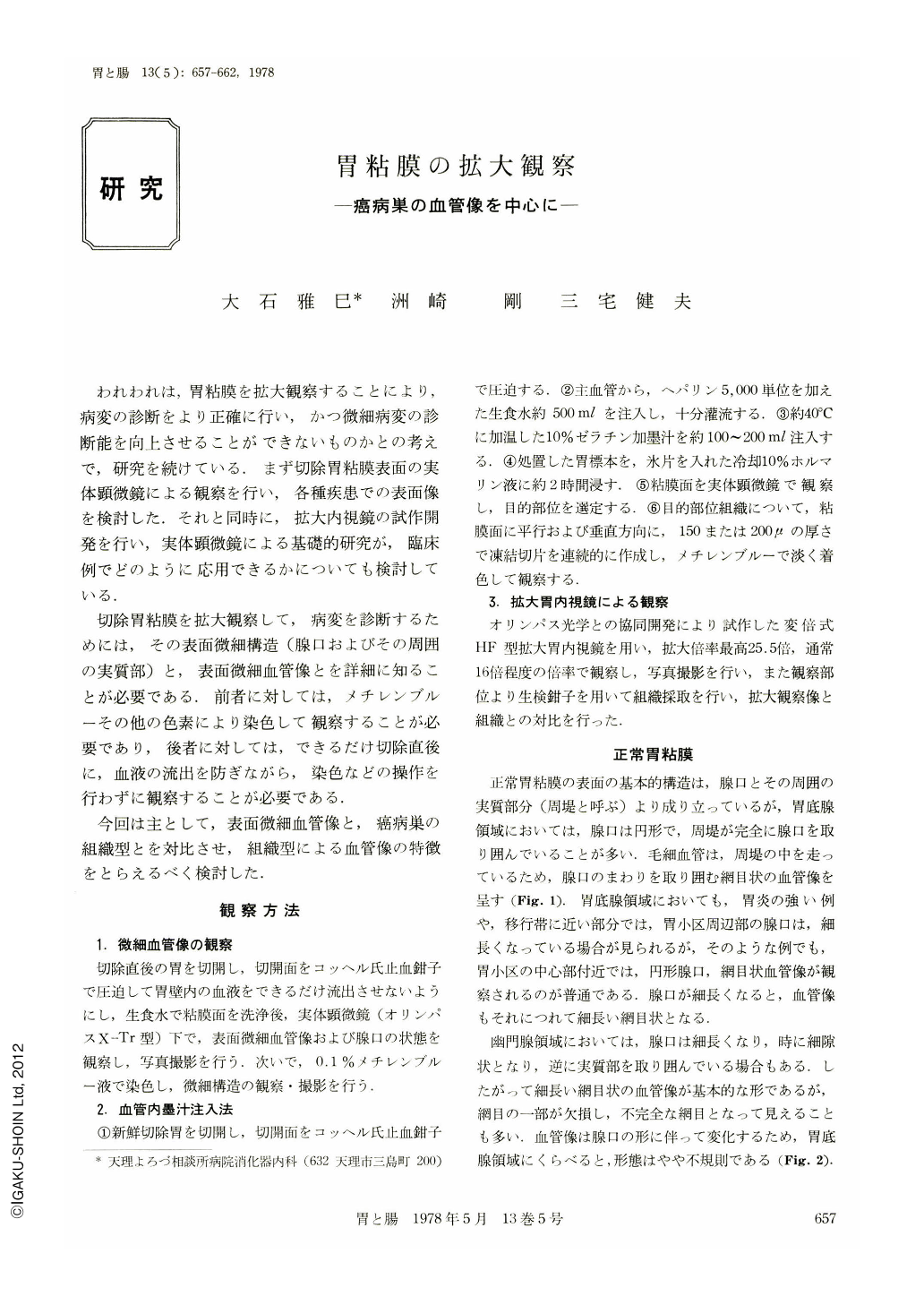
われわれは,胃粘膜を拡大観察することにより,病変の診断をより正確に行い,かつ微細病変の診断能を向上させることができないものかとの考えで,研究を続けている.まず切除胃粘膜表面の実体顕微鏡による観察を行い,各種疾患での表面像を検討した.それと同時に,拡大内視鏡の試作開発を行い,実体顕微鏡による基礎的研究が,臨床例でどのように応用できるかについても検討している.
切除胃粘膜を拡大観察して,病変を診断するためには,その表面微細構造(腺口およびその周囲の実質部)と,表面微細血管像とを詳細に知ることが必要である.前者に対しては,メチレンブルーその他の色素により染色して観察することが必要であり,後者に対しては,できるだけ切除直後に,血液の流出を防ぎながら,染色などの操作を行わずに観察することが必要である.
The authors attempted to observe the fine structure and microangiographic features of the gastric mucosa by a dissecting microscope as the basic study in order to make diagnosis. The magnification endoscopy (magnifiable HF Type) which we developed was clinically applied. Observations on the fine structure and angiography of the mucosa were disclosed to be very useful in order to make correct diagnosis.
The results appear to reveal that the microangiographic examination of the superficial mucosa made possible the differentiation between malignancy and benignity, and even an estimation of histological type of cancer.
The basic structure in the normal mucosa is seen at the gastric glandular openning and its surroundings. The openning are round in the fundic region and longitudinal or slit-like in the pyloric region. Microangiographic features are reticular and round in the former and elongated, reticular but partly interrupted in the latter.
The surface structure of cancer lesion and angiographic features are generally characterized by an irregularity in their shape, distribution and arrangement.
As characteristic features in individual histological type of cancer, a reticular pattern in angiograpy is often seen in well differentiated cancer of depressed type. In well-differentiated cancer of protruded type, the parallel running blood vessels are arranged in waves. In cases of papillary adenocarcinoma showing a large solid protrusion, a racemose pattern is observed in the lateral view of the blood vessels. In anaplastic cancer, the vessels are characterized by an irregular, uneven arborizing or short linear pattern.
Copyright © 1978, Igaku-Shoin Ltd. All rights reserved.