Japanese
English
- 有料閲覧
PPV購入案内
1,320円 (1,200円+税10%) こちらは、1674ページから1678ページの文献です。購入には医書.jp本会員登録が必要です。
会員登録完了後に改めて上記「購入サイトへ」を選択してください。
または「購入サイトへ」選択後に「購入ログイン」、「新規会員登録」の順に進んでください。
会員登録について詳しくはをご確認ください。
- Abstract 文献概要
- 1ページ目 Look Inside
- 参考文献 Reference
- サイト内被引用 Cited by
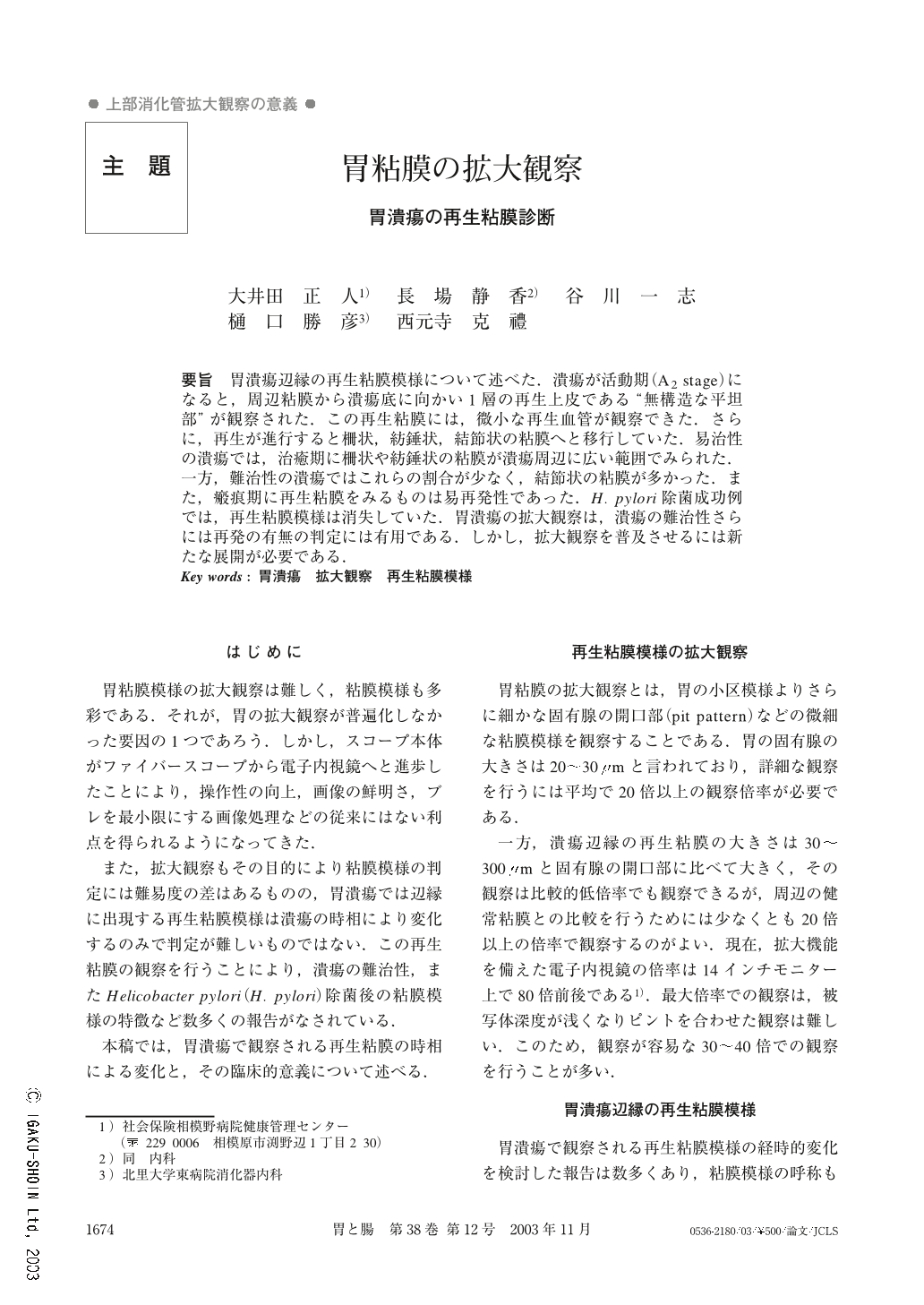
要旨 胃潰瘍辺縁の再生粘膜模様について述べた.潰瘍が活動期(A2 stage)になると,周辺粘膜から潰瘍底に向かい1層の再生上皮である“無構造な平坦部”が観察された.この再生粘膜には,微小な再生血管が観察できた.さらに,再生が進行すると柵状,紡錘状,結節状の粘膜へと移行していた.易治性の潰瘍では,治癒期に柵状や紡錘状の粘膜が潰瘍周辺に広い範囲でみられた.一方,難治性の潰瘍ではこれらの割合が少なく,結節状の粘膜が多かった.また,瘢痕期に再生粘膜をみるものは易再発性であった.H. pylori除菌成功例では,再生粘膜模様は消失していた.胃潰瘍の拡大観察は,潰瘍の難治性さらには再発の有無の判定には有用である.しかし,拡大観察を普及させるには新たな展開が必要である.
We described regenerative epithelium during the healing process of chronic gastric ulcer. In the A2 stage, flattened mucosal pattern could be observed intially at the margin of the ulcer. As the regenerative changes advanced, the epithelium developed a palisade-like, spindle-like and nodule-like appearance. In nodule-like epithelia, histological findings showed mature regeration with pseudo pyloric glands. After that, nodule-like epithelium developed a normal mucosal pattern.
Magnifying endoscopic findings made it possible to evaluate the intractability of healing and to predict relapse of the ulcer.
In conclusion, magnifying endoscopy enables us to easily estimate ulcer healing and relapse.
Copyright © 2003, Igaku-Shoin Ltd. All rights reserved.