Japanese
English
- 有料閲覧
PPV購入案内
1,320円 (1,200円+税10%) こちらは、1625ページから1628ページの文献です。購入には医書.jp本会員登録が必要です。
会員登録完了後に改めて上記「購入サイトへ」を選択してください。
または「購入サイトへ」選択後に「購入ログイン」、「新規会員登録」の順に進んでください。
会員登録について詳しくはをご確認ください。
- Abstract 文献概要
- 1ページ目 Look Inside
- 参考文献 Reference
- サイト内被引用 Cited by
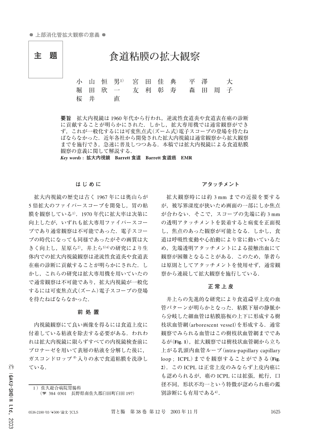
要旨 拡大内視鏡は1960年代から行われ,逆流性食道炎や食道表在癌の診断に貢献することが明らかにされた.しかし,拡大専用機では通常観察ができず,これが一般化するには可変焦点式(ズーム式)電子スコープの登場を待たねばならなかった.近年各社から開発された拡大内視鏡は通常観察から拡大観察までを施行でき,急速に普及しつつある.本稿では拡大内視鏡による食道粘膜観察の意義に関して解説する.
Magnification endoscopy is useful for the diagnosis of esophageal diseases. Some endoscopists have reported the usefulness of magnification endoscopy for the diagnosis of reflux esophagitis, superficial squamous cell cancer and Barrett's esophagus 1)~5).
The focus range of magnification endoscopy is narrow, so a transparent attachment is useful to keep the endoscope at an adequate distance from the lesion. However, sometimes the transparent attachment causes contact bleeding, so usually we perform magnification endoscopy without the attachment.
Normal endoscopy can show submucosal vessels and arborescent vessels, but magnification endoscopy can show intra-papillary capillary loop (ICPL). ICPL of superficial cancer is different from that of the normal esophagus.
Magnification endoscopy is also useful in the diagnosis of superficial Barrett's esophageal cancer. Normal endoscopy shows a shallow depressed lesion (Fig. 9), but magnification endoscopy shows irregular vascular pattern and no pit pattern (Fig. 10). Therefore, magnification endoscopy is useful for the diagnosis of esophageal disease.
Copyright © 2003, Igaku-Shoin Ltd. All rights reserved.