Japanese
English
- 有料閲覧
- Abstract 文献概要
- 1ページ目 Look Inside
大腸粘膜表面に解剖学上無名の溝(innominate grooves)があることを指摘し,それがX線像上にnetwork pattern,spiculationおよびdotted patternとして現われることがあるのを最初に報告したのはWilliams1)である.しかし事実の指摘に終わり診断的意味づけは行われていない.ついで狩谷ら2)3)は実体顕微鏡,切除大腸の二重造影像にてこのnetwork patternがX線像における表現可能な最小単位で大腸二重造影像の基本像であり,炎症性疾患,ことに潰瘍性大腸炎のX線所見による治癒判定には欠くことのできぬものであることを指摘した.われわれはより微細なX線像を得ることを目的にして1972年より微小管球を用いて切除大腸の拡大撮影を行い,このnetwork patternの微細構造の詳細な検討を行い報告してきた4)5).またこれと並行して拡大撮影の臨床への応用も行ってきた6).本稿では臨床の場における拡大撮影による大腸粘膜の微細診断の意義について述べる.
研究方法および目的
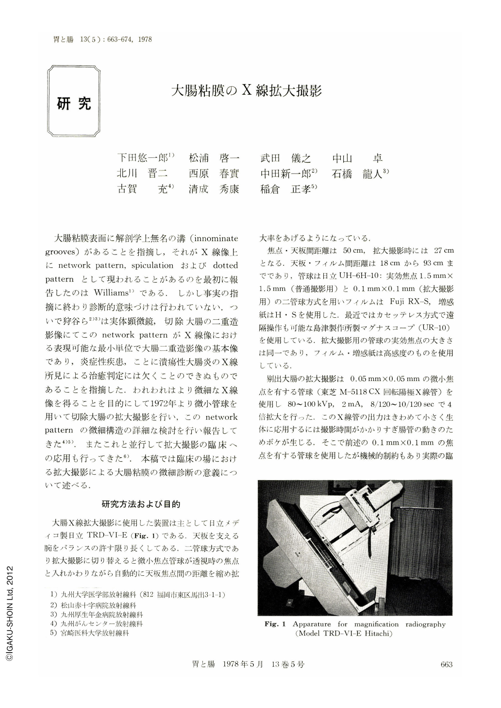
大腸X線拡大撮影に使用した装置は主として目立メディコ製日立TRD-VI-E(Fig. 1)である.天板を支える腕をバランスの許す限り長くしてある.二管球方式であり拡大撮影に切り替えると微小焦点管球が透視時の焦点と入れかわりながら自動的に天板焦点問の距離を縮め拡大率をあげるようになっている.
We compared direct magnification radiographies of the colon using a fluoroscopy unit having a microfocus tube with the conventional radiographies and obtained the following results.
1) Magnification radiography was superior to the conventional technique in delineating the marginal patterns such as spiculation in the normal mucosa and small niches in ulcerative colitis.
2) There was no difference in delineating the double contrast mucosal pattern en face between the magnification and conventional technique.
3) Considering motion of the colon, function of the unit, and patient's exposure dosage, three times magnification is probably the maximum limit.
4) This technique need not be done on every case, but should be performed on selected cases where its advantage is expected.

Copyright © 1978, Igaku-Shoin Ltd. All rights reserved.


