Japanese
English
- 有料閲覧
PPV購入案内
1,320円 (1,200円+税10%) こちらは、387ページから397ページの文献です。購入には医書.jp本会員登録が必要です。
会員登録完了後に改めて上記「購入サイトへ」を選択してください。
または「購入サイトへ」選択後に「購入ログイン」、「新規会員登録」の順に進んでください。
会員登録について詳しくはをご確認ください。
- Abstract 文献概要
- 1ページ目 Look Inside
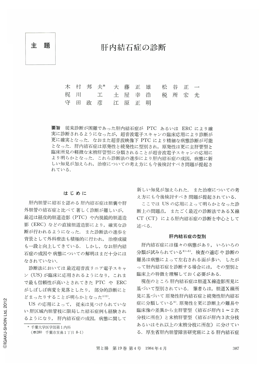
要旨 従来診断が困難であった肝内結石症がPTCあるいはERCにより確実に診断されるようになったが,超音波電子スキャンの臨床応用により診断が更に確実となった.なおまた超音波映像下PTCにより精細な病態診断が可能となった.肝内結石症は原発性と続発性に型別され,原発性は更に主肝管型と臨床所見の軽微な末梢肝管型に分類されることが超音波電子スキャンの応用により明らかとなった.これら診断法の進歩により肝内結石症の成因,病態に新しい知見が加えられ,治療についての考え方にも今後検討すべき問題が提起されている.
Precise diagnosis of hepatolithiasis before surgery has become possible by recent advance of diagnostic modalities for direct cholangiography such as percutaneous transhepatic cholangiography (PTC) and endoscopic retrograde cholangiography (ERC). Furthermore, it has been clarified by the development of real-time ultrasound (US) that both of conventional PTC and ERC have difficulty in visualizing the entire feature of the intrahepatic biliary tree in patients with stenotic portion at the distal end of the segmentally dilated bile duct containing stones.
The purpose of this paper was to assess the current diagnosis of hepatolithiasis based on our classification. According to our cholangiographic study, this condition was divided into two groups, primary (Fig. 1) and secondary (Fig. 2). The clinical and cholangiographic characteristics of each group were summarized in Table 1. According to the location of intrahepatic gallstones with associated segmental dilatation of the intrahepatic bile ducts, primary hepatolithiasis was divided into two subgroups: the type of the major intrahepatic bile duct (Figs. 4 and 5) in which the lesion was locating predominantly in the major hepatic ducts, and the type of peripheral intrahepatic bile ducts (Fig. 7) in which the lesion was distributed only in the smaller branches of the hepatic ducts at the third order branches or more proximal. The rate of incidence of the type of peripheral intrahepatic bile ducts, which had the mildest clinical features (Tables 3 and 4), has remarkably increased after the introduction of real-time US.
The diagnostic capability of various modalities in detecting intrahepatic gallstones was studied in 28 patients (Table 6). Deffinite diagnosis of intrahepatic gallstones was made in 46.5% by PTC and/or ERC, 96.4% by US, and 100% by US-guided PTC, respectively.
It was concluded that US was the most reliable modality in screening intrahepatic gallstones, and that US-guided PTC was the most accurate procedure to give the precise diagnosis in patients with insufficient informations by ERC (Fig. 7).
Copyright © 1984, Igaku-Shoin Ltd. All rights reserved.