Japanese
English
- 有料閲覧
PPV購入案内
1,320円 (1,200円+税10%) こちらは、1299ページから1302ページの文献です。購入には医書.jp本会員登録が必要です。
会員登録完了後に改めて上記「購入サイトへ」を選択してください。
または「購入サイトへ」選択後に「購入ログイン」、「新規会員登録」の順に進んでください。
会員登録について詳しくはをご確認ください。
- Abstract 文献概要
- 1ページ目 Look Inside
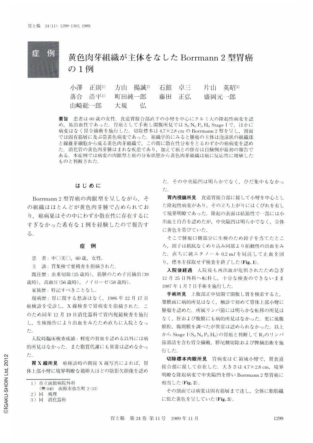
要旨 患者は60歳の女性.食道胃接合部直下の小彎を中心にクルミ大の隆起性病変を認め,易出血性であった.胃癌として手術し開腹所見ではS0N0P0H0 Stage Ⅰで,ほかに病変はなく胃全摘術を施行した.切除標本は4.7×2.8cmのBorrmann 2型を呈し,割面では固有筋層に及ぶ帯黄色病変であった.組織学的にみると腫瘍の主体は泡沫状の組織球と線維芽細胞から成る黄色肉芽組織で,この間に散在性分布をとるわずかの癌病変を認めた.消化管の黄色肉芽腫はまれな疾患であり,加えて癌との併存は自験例が最初の報告である.本症例では病変の肉眼型と癌の分布状態から黄色肉芽組織は癌に反応性に増殖したものと判断された.
A 60-year-old housewife was examined for gastric cancer without any symptoms.
By roentgenography and endoscopy, a protruding mass on the lesser curvature near the esophagogastric junction was revealed and diagnosed as cancer (Fig. 1). Total gastrectomy with distal pancreatectomy and splenectomy was performed on Jan. 7, 1987. Resected specimen showed macroscopically a protruding, well delimited mass with central ulceration (4.7 × 2.8 cm in size) on the mucosa (Fig. 2) and yellowish solid lesion involving mucosa and proper muscular layer on the cut surface (Fig. 3). However, histological examination disclosed that the lesion consisted, for the most part, of histiocytes with foamy cytoplasm and fibroblasts (Fig. 4), and only a few small foci of carcinomas with lymphocyte infiltration were scattered in the xanthogranuloma tissue (Figs. 6 and 7).
This is believed to be the first recorded instance of xanthogranuloma accompanying carcinoma in the same tumor of the stomach.
Copyright © 1989, Igaku-Shoin Ltd. All rights reserved.