Japanese
English
- 有料閲覧
- Abstract 文献概要
- 1ページ目 Look Inside
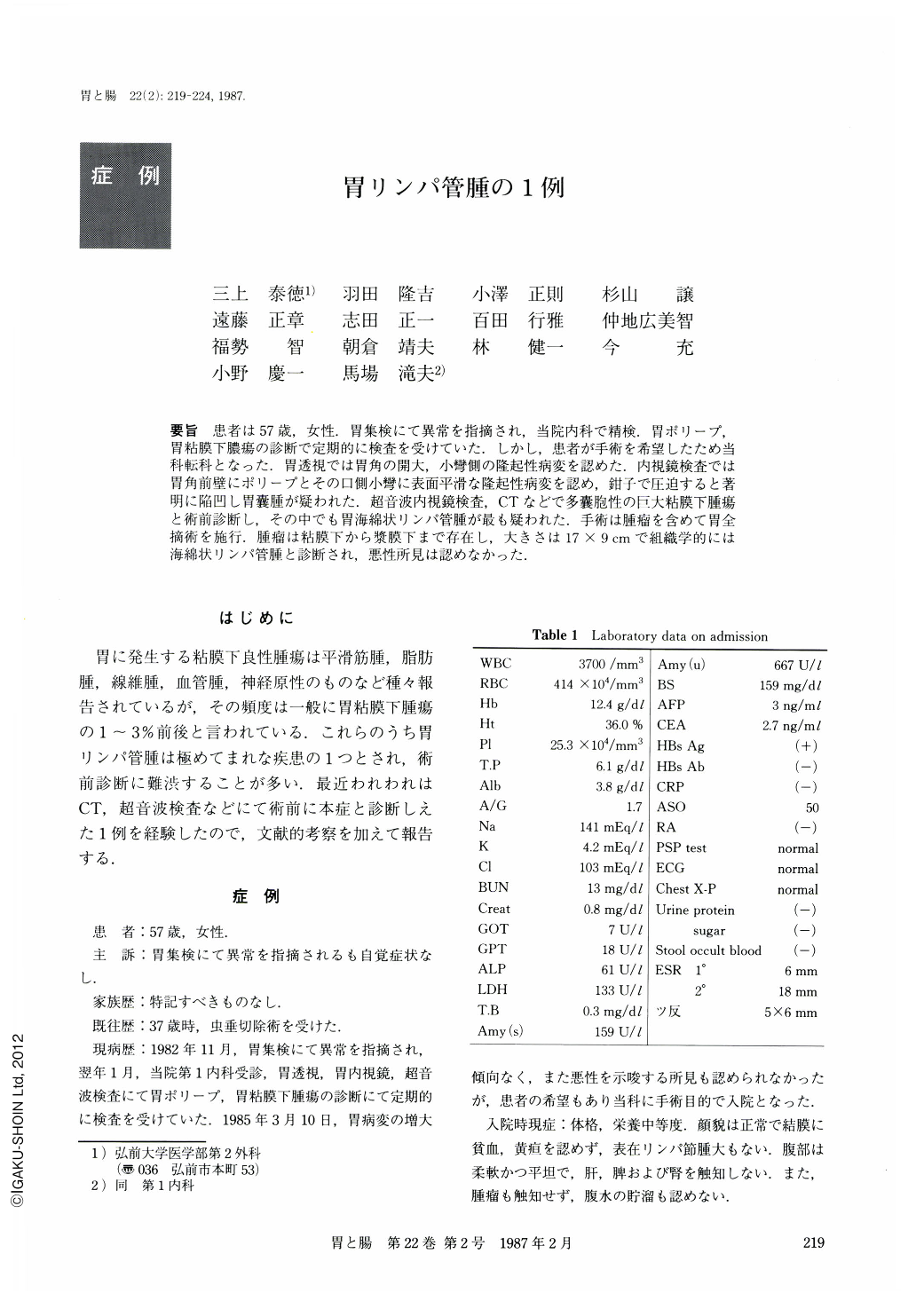
要旨 患者は57歳,女性.胃集検にて異常を指摘され,当院内科で精検.胃ポリープ,胃粘膜下膿瘍の診断で定期的に検査を受けていた.しかし,患者が手術を希望したため当科転科となった.胃透視では胃角の開大,小彎側の隆起性病変を認めた.内視鏡検査では胃角前壁にポリープとその口側小彎に表面平滑な隆起性病変を認め,鉗子で圧迫すると著明に陥凹し胃囊腫が疑われた.超音波内視鏡検査,CTなどで多囊胞性の巨大粘膜下腫瘍と術前診断し,その中でも胃海綿状リンパ管腫が最も疑われた.手術は腫瘤を含めて胃全摘術を施行.腫瘤は粘膜下から漿膜下まで存在し,大きさは17×9cmで組織学的には海綿状リンパ管腫と診断され,悪性所見は認めなかった.
A case of cavernous lymphangioma of the stomach, which developed in the lesser curvature, is reported. In addition, literature review was made, which identified 45 cases reported so far in Japan. Six cases of giant (150 mm) lymphangioma are reviewed as well (Table 2).
In November 1982, 57-year old asymptomatic female was suspected of having a submucosal tumor of the stomach at a gastric carcinoma mass survey. X-ray examination revealed a giant submucosal tumor in the lesser curvature (Fig. 1). Gastrofiberscopy, however, showed multiple protuberant lesions. The surfaces were smooth and similar to the surrounding normal mucosa. The mucosa seemed to have fluctuation as well as bridging folds (Fig. 2). Normal five layers of gastric wall were not visualized by endoscopic ultrasonography (EUS ), which highly suggests submucosal multilocular lesions. Computed tomography (CT) demonstrated a large low-density tumor extending from the lesser curvature to the hilus of the spleen (Fig. 4). The tumor had no signs of malignancy on CT. In April 1985, total gastrectomy and tumor resection were performed. Resected specimen showed a giant submucosal and subserous tumor, measuring 16 × 9 × 6 cm in size (Fig. 5). The cut surface was multilocular like honeycomb with serous material in the cysts (Fig. 6 a). Histologically, the wall consisted of a single layer of endothelium and there was eosinophilic material in the cyst. Final histo-logical diagnosis was cavernous lymphangioma of the stomach (Fig. 6 b). Postoperative course was uneventful.

Copyright © 1987, Igaku-Shoin Ltd. All rights reserved.


