Japanese
English
- 有料閲覧
PPV購入案内
1,320円 (1,200円+税10%) こちらは、685ページから690ページの文献です。購入には医書.jp本会員登録が必要です。
会員登録完了後に改めて上記「購入サイトへ」を選択してください。
または「購入サイトへ」選択後に「購入ログイン」、「新規会員登録」の順に進んでください。
会員登録について詳しくはをご確認ください。
- Abstract 文献概要
- 1ページ目 Look Inside
- サイト内被引用 Cited by
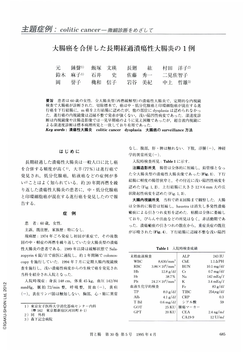
要旨 患者は60歳の女性.全大腸炎型(再燃緩解型)の潰瘍性大腸炎で,定期的な内視鏡検査で大腸癌が診断された.切除標本で,癌は中・低分化腺癌と印環細胞癌が混在する進行癌を下行結腸に,m癌を上行結腸に認めたが,他の部位にdysplasiaは認められなかった.進行癌の内視鏡像は辺縁不整で発赤が強くない,浅い陥凹性病変であった.深達度診断は内視鏡像や注腸造影像では一見早期癌のように見え困難であったが,超音波内視鏡による深達度診断は標本病理所見と一致しており有用であった.
A 60-year-old woman was admitted to our hospital in December, 1994, for examination and treatment of colitic cancer detected in another hospital during a periodical endoscopic examination. Barium enema showed disappearance of the haustra and shortening of the entire colon (type of total colitis). Recurrence and remission had occared during the previous 20 years. Histological findings disclosed moderately and poorly differentiated tubular adenocarcinoma mixed with sig-net-ring cell carcinoma invading as far as the serosa of the descending colon and, well differentiated tubular adenocarcinoma limited to the mucosa of the ascending colon. Macroscopically, the descending colonic cancer was a slightly reddish and excavated lesion with irregular margin. It was very difficult to diagnose the depth of invasion of the cancer (similar to early colonic cancer) in the colonoscopic and roentgenographic findings in this case, but endoscopic ultrasonography was very useful for this purpose.
Copyright © 1995, Igaku-Shoin Ltd. All rights reserved.