Japanese
English
- 有料閲覧
PPV購入案内
1,320円 (1,200円+税10%) こちらは、1159ページから1165ページの文献です。購入には医書.jp本会員登録が必要です。
会員登録完了後に改めて上記「購入サイトへ」を選択してください。
または「購入サイトへ」選択後に「購入ログイン」、「新規会員登録」の順に進んでください。
会員登録について詳しくはをご確認ください。
- Abstract 文献概要
- 1ページ目 Look Inside
- サイト内被引用 Cited by
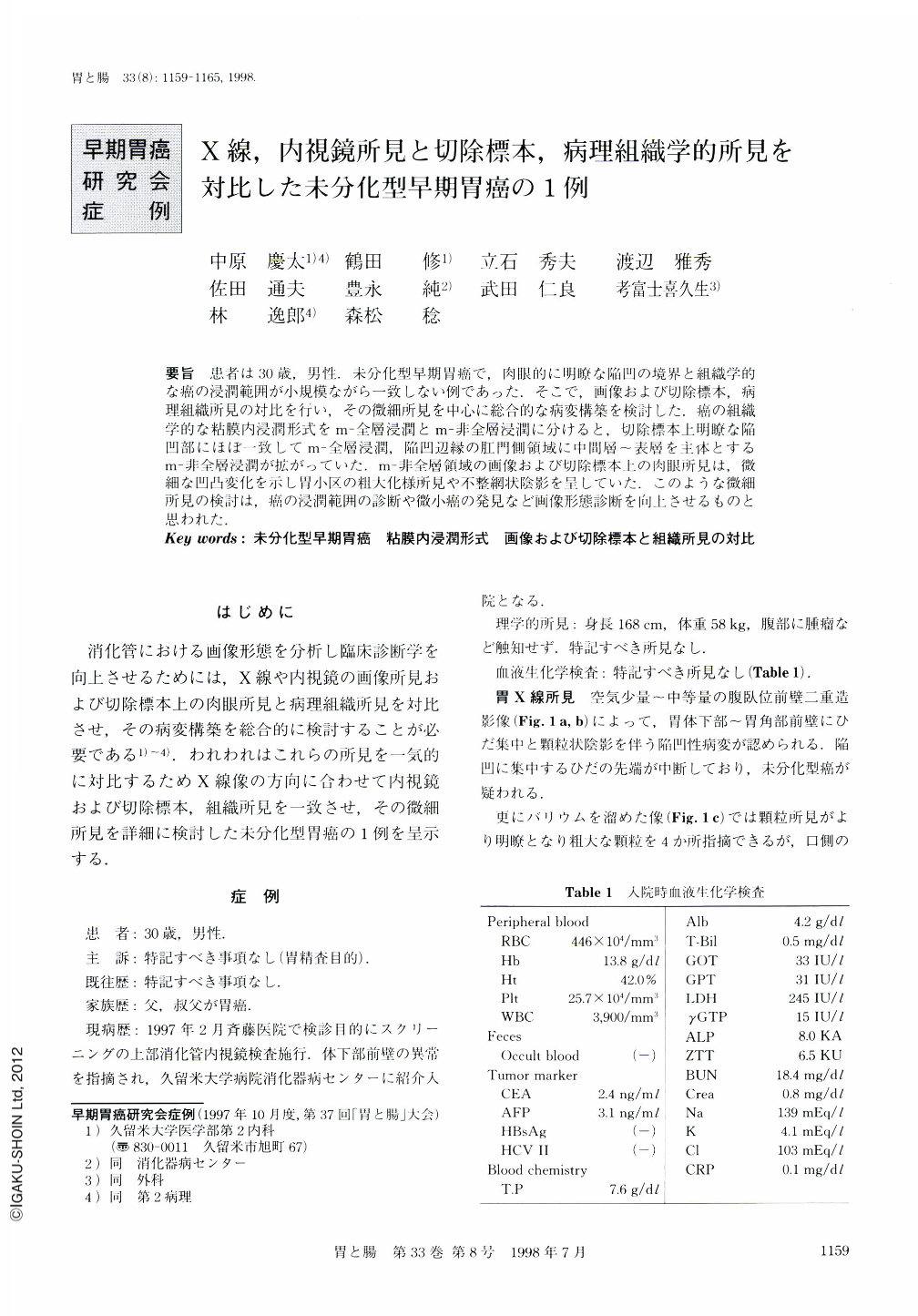
要旨 患者は30歳,男性.未分化型早期胃癌で,肉眼的に明瞭な陥凹の境界と組織学的な癌の浸潤範囲が小規模ながら一致しない例であった.そこで,画像および切除標本,病理組織所見の対比を行い,その微細所見を中心に総合的な病変構築を検討した.癌の組織学的な粘膜内浸潤形式をm-全層浸潤とm-非全層浸潤に分けると,切除標本上明瞭な陥凹部にほぼ一致してm-全層浸潤,陥凹辺縁の肛門側領域に中間層~表層を主体とするm-非全層浸潤が拡がっていた.m-非全層領域の画像および切除標本上の肉眼所見は,微細な凹凸変化を示し胃小区の粗大化様所見や不整網状陰影を呈していた.このような微細所見の検討は,癌の浸潤範囲の診断や微小癌の発見など画像形態診断を向上させるものと思われた.
An undifferentiated type of early gastric cancer in a 30-year-old man was investigated because of the discrepancy between macroscopic and histological diagnosis in the lesional extension. Macroscopic findings of clear depression of the lesional margin corresponded approximately with the area of total mucosal layer invasion. However, macroscopic findings of roughly elevated granular or irregular and reticular pattern or uneven mucosal appearance corresponded approximately with the area where there was half invasion of the mucosal layer. We believe that careful evalution of radiographic and endoscopic findings of fine mucosal patterns could be of half to detect and correctly diagnose minute gastric cancer.
Copyright © 1998, Igaku-Shoin Ltd. All rights reserved.