Japanese
English
- 有料閲覧
PPV購入案内
1,320円 (1,200円+税10%) こちらは、1109ページから1114ページの文献です。購入には医書.jp本会員登録が必要です。
会員登録完了後に改めて上記「購入サイトへ」を選択してください。
または「購入サイトへ」選択後に「購入ログイン」、「新規会員登録」の順に進んでください。
会員登録について詳しくはをご確認ください。
- Abstract 文献概要
- 1ページ目 Look Inside
- サイト内被引用 Cited by
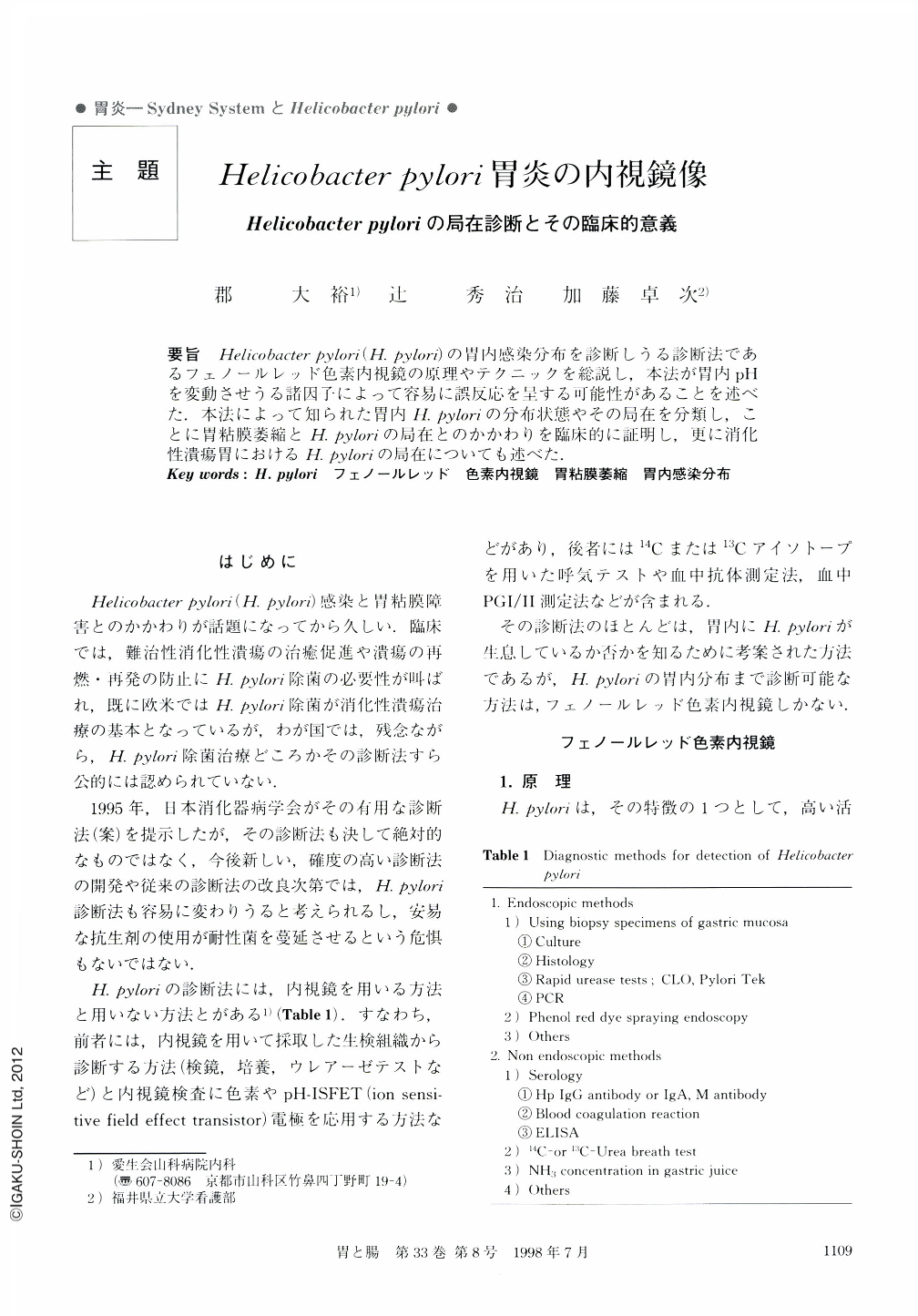
要旨 Helicobacter pylori(H. Pylori)の胃内感染分布を診断しうる診断法であるフェノールレッド色素内視鏡の原理やテクニックを総説し,本法が胃内pHを変動させうる諸因子によって容易に誤反応を呈する可能性があることを述べた.本法によって知られた胃内H. Pyloriの分布状態やその局在を分類し,ことに胃粘膜萎縮とH. Pyloriの局在とのかかわりを臨床的に証明し,更に消化性潰瘍胃におけるH. Pyloriの局在についても述べた.
The principle of Phenol red dye spraying endoscopy and its techniques are explained, showing H. pylori localization in human gastric mucosa as a red-color region. The red-color region with phenol red solution is classified into four types; diffusely stained type, regionally stained type, patchily stained type and the unstained type. According to the classification of the fun-dic-pyloric border by Kimura T and Takemoto T, the unstained type is mainly discernible in the C0 pattern, and the regionally or diffusely stained types increase with the oral shift of the border. In the O3 pattern, the unstained type is dominant. From these data, it can be said that gastric mucosal atrophy is most likely to be associated with continuous infection by H. pylori. In addition, a red color region is always visible at the margins of gastric ulcers, diffusely or over the whole circumference.
Copyright © 1998, Igaku-Shoin Ltd. All rights reserved.