Japanese
English
- 有料閲覧
- Abstract 文献概要
- 1ページ目 Look Inside
- サイト内被引用 Cited by
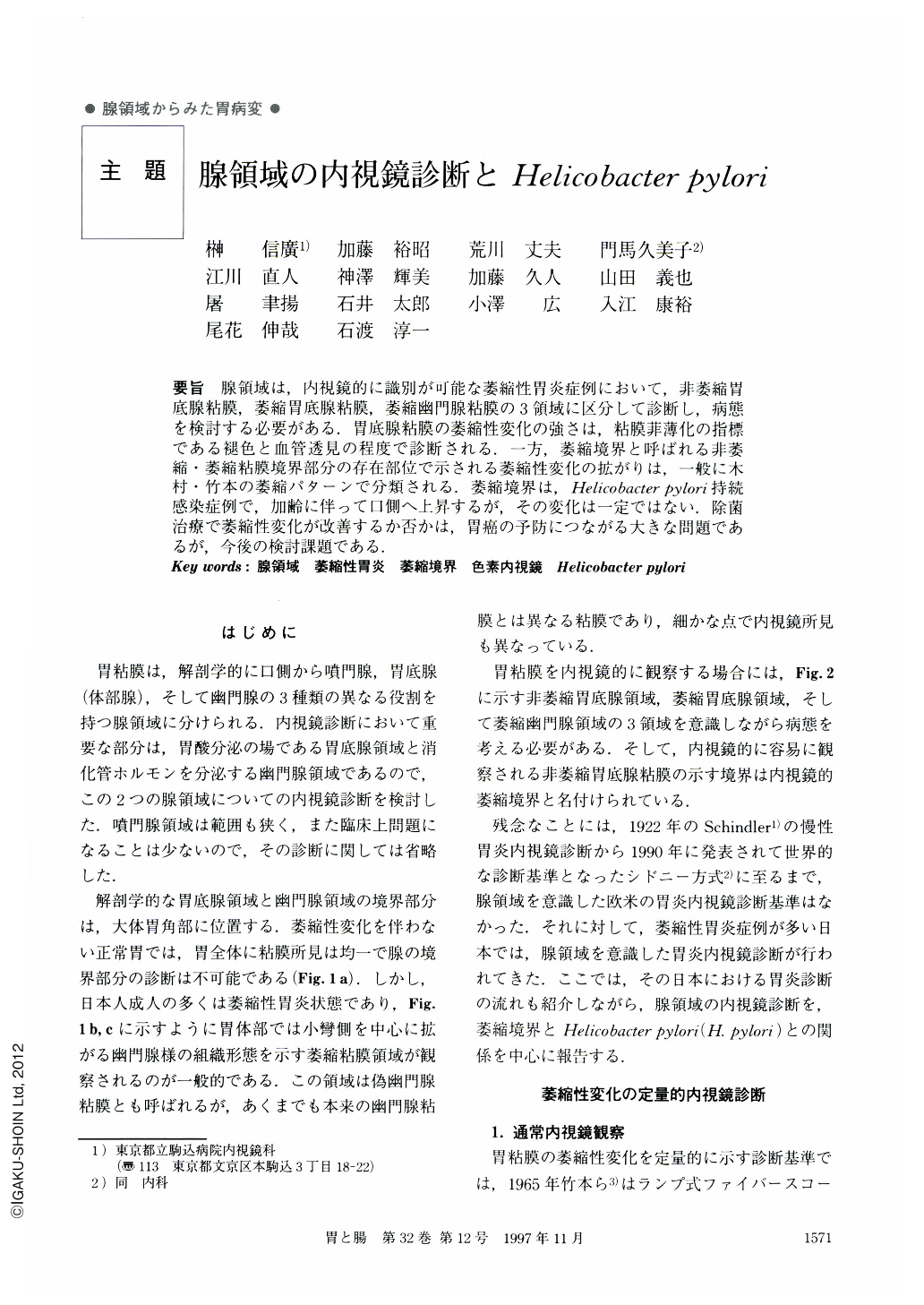
要旨 腺領域は,内視鏡的に識別が可能な萎縮性胃炎症例において,非萎縮胃底腺粘膜,萎縮胃底腺粘膜,萎縮幽門腺粘膜の3領域に区分して診断し,病態を検討する必要がある.胃底腺粘膜の萎縮性変化の強さは,粘膜菲薄化の指標である褪色と血管透見の程度で診断される.一方,萎縮境界と呼ばれる非萎縮・萎縮粘膜境界部分の存在部位で示される萎縮性変化の拡がりは,一般に木村・竹本の萎縮パターンで分類される.萎縮境界は,elicobacter Pylori持続感染症例で,加齢に伴って口側へ上昇するが,その変化は一定ではない.除菌治療で萎縮性変化が改善するか否かは,胃癌の予防につながる大きな問題であるが,今後の検討課題である.
An endoscopic diagnosis of gastric glandular areas is valuable in patients with atrophic gastritis whose gastric mucosa is able to divide into three areas; a nonatrophic fundic area, an atrophic fundic area and an atrophic pyloric mucosa. The severities of the atrophic changes are assessed by a discoloration and a transparency of blood vessels which indicate a thinning of the mucosa. On the other hand, the extensions of atrophic change are demonstrated by atrophic borders between a non-atrophic area and an atrophic area and are classified according to the norms of Kimura and Takemoto. The cephalod shifts of atrophic borders are observed in cases with Helicobacter pylori positive chronic active gastritis. However, those changes are discontinuous and occasionally advance rapidly. Endoscopic studies assessing the effects of Helicobacter pylori eradication on regression/progression of atrophic change of the gastric mucosa and subsequent development of gastric cancer are needed.

Copyright © 1997, Igaku-Shoin Ltd. All rights reserved.


