Japanese
English
- 有料閲覧
PPV購入案内
1,320円 (1,200円+税10%) こちらは、545ページから551ページの文献です。購入には医書.jp本会員登録が必要です。
会員登録完了後に改めて上記「購入サイトへ」を選択してください。
または「購入サイトへ」選択後に「購入ログイン」、「新規会員登録」の順に進んでください。
会員登録について詳しくはをご確認ください。
- Abstract 文献概要
- 1ページ目 Look Inside
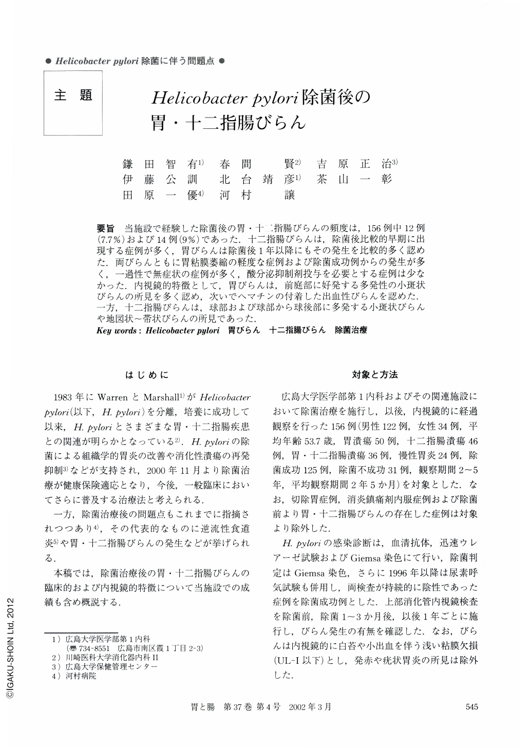
要旨 当施設で経験した除菌後の胃・十二指腸びらんの頻度は,156例中12例(7.7%)および14例(9%)であった.十二指腸びらんは,除菌後比較的早期に出現する症例が多く,胃びらんは除菌後1年以降にもその発生を比較的多く認めた.両びらんともに胃粘膜萎縮の軽度な症例および除菌成功例からの発生が多く,一過性で無症状の症例が多く,酸分泌抑制剤投与を必要とする症例は少なかった.内視鏡的特徴として,胃びらんは,前庭部に好発する多発性の小斑状びらんの所見を多く認め,次いでヘマチンの付着した出血性びらんを認めた.一方,十二指腸びらんは,球部および球部から球後部に多発する小斑状びらんや地図状~帯状びらんの所見であった.
Out of 156 cases of patients treated for Helicobacter pylori (H. pylori), there were 12 examples of gastric erosions (7.7%) and 14 examples of duodenal erosions (9%).
As clinical features, the duodenal erosions were mainly recognized 1~3 months after H. pylori eradication. On the other hand, the gastric erosions were mainly recognized 1 year after H. pylori eradication. Both types of erosions were mainly recognized from mild atrophy of the gastric mucosa in spite of successful H. pylori eradication. These were transient lesions which disappeared after follow-up in most case. There were also many subclinical cases, but only a few of them required the administration of acid suppressives.
As endoscopic features, the gastric erosions were mainly recognized as erosions consisting of multiple small spots and hemorrhagic erosions. On the other hand, the duodenal erosions were mainly recognized as a comparatively large map-like state produced by banded erosions.
Copyright © 2002, Igaku-Shoin Ltd. All rights reserved.