Japanese
English
- 有料閲覧
PPV購入案内
1,320円 (1,200円+税10%) こちらは、671ページから682ページの文献です。購入には医書.jp本会員登録が必要です。
会員登録完了後に改めて上記「購入サイトへ」を選択してください。
または「購入サイトへ」選択後に「購入ログイン」、「新規会員登録」の順に進んでください。
会員登録について詳しくはをご確認ください。
- Abstract 文献概要
- 1ページ目 Look Inside
- サイト内被引用 Cited by
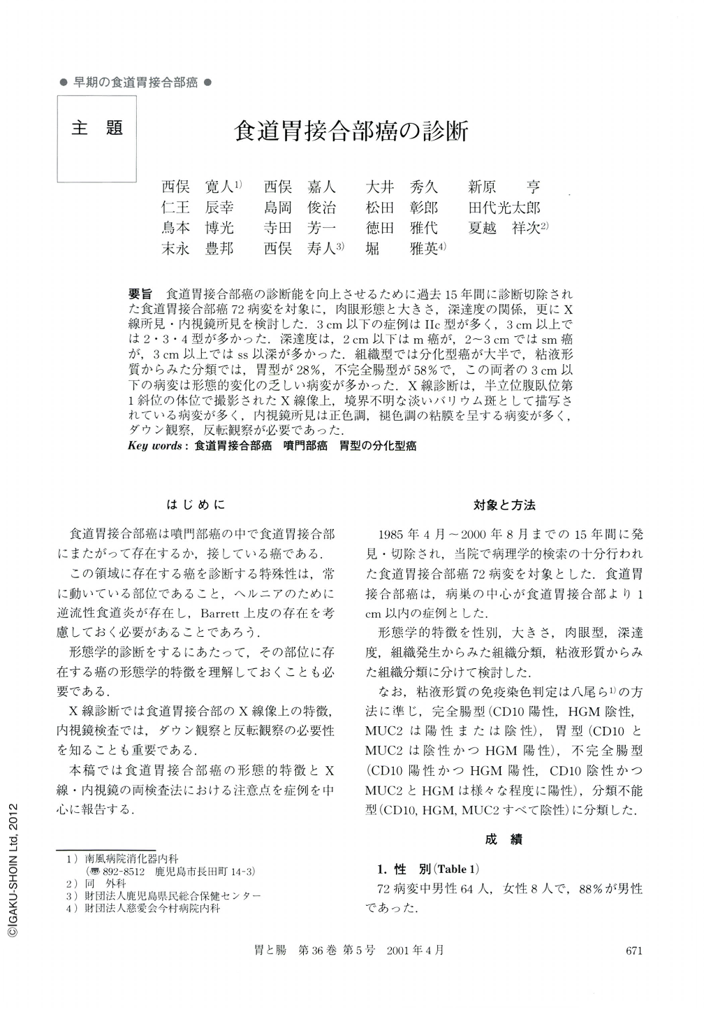
要旨 食道胃接合部癌の診断能を向上させるために過去15年間に診断切除された食道胃接合部癌72病変を対象に,肉眼形態と大きさ,深達度の関係,更にX線所見・内視鏡所見を検討した.3cm以下の症例はⅡc型が多く,3cm以上では2・3・4型が多かった.深達度は,2cm以下はm癌が,2~3cmではsm癌が,3cm以上ではss以深が多かった.組織型では分化型癌が大半で,粘液形質からみた分類では,胃型が28%,不完全腸型が58%で,この両者の3cm以下の病変は形態的変化の乏しい病変が多かった.X線診断は,半立位腹臥位第1斜位の体位で撮影されたX線像上,境界不明な淡いバリウム斑として描写されている病変が多く,内視鏡所見は正色調,褪色調の粘膜を呈する病変が多く,ダウン観察,反転観察が必要であった.
To improve diagnostic ability of the cancer of the cardioesophageal junction, 72 lesions of the cardioesophageal cancer diagnosed and resected last 15 years were analyzed on the macroscopic classification, size, dephth of invasion, and the radiological and endoscopic findings. Many of the lesions less than 3 cm in size were type IIc, whereas the lesions more than 3 cm in size were tend to be type 2, 3, 4 in macroscopic shape. The relationship between size and the depth of invasion were summarized as follows : the lesion less than 2 cm in size was may be limited in m invasion, the lesion two to three cm in size may have sm invasion and the lesion more than 3 cm in size may have ss or deeper invasion. Most of the lesions were the differentiated type of the histological classification. From the viewpoint of mucinous type classification, 28% of the entire lesion was the stomach type and 58% was the incomplete intestinal type, the lesion of both types less than 3 cm in size did not have prominent macroscopic changes.
From the radiological diagnostic viewpoints, many lesions were detected as illdemarcated pale barium patches on the film exposed by the semi-standing, prone and first oblique position. Endoscopically, they were normal or brownish colored mucosa detected by down and turn-over views.
Copyright © 2001, Igaku-Shoin Ltd. All rights reserved.