Japanese
English
- 有料閲覧
- Abstract 文献概要
- 1ページ目 Look Inside
- 参考文献 Reference
- サイト内被引用 Cited by
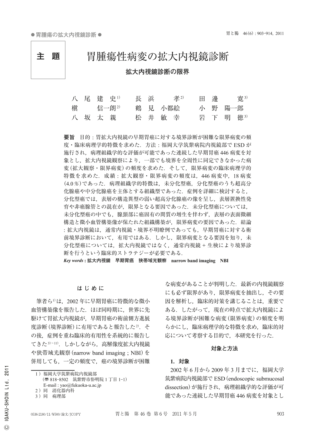
要旨 目的:胃拡大内視鏡の早期胃癌に対する境界診断が困難な限界病変の頻度・臨床病理学的特徴を求めた.方法:福岡大学筑紫病院内視鏡部でESDが施行され,病理組織学的な評価が可能であった連続した早期胃癌446病変を対象とし,拡大内視鏡観察により,一部でも境界を全周性に同定できなかった病変(拡大観察・限界病変)の頻度を求めた.そして,限界病変の臨床病理学的特徴を求めた.成績:拡大観察・限界病変の頻度は,446病変中,18病変(4.0%)であった.病理組織学的特徴は,未分化型癌,分化型癌のうち超高分化腺癌や中分化腺癌を主体とする組織型であった.症例を詳細に検討すると,分化型癌では,表層の構造異型の弱い超高分化腺癌の像を呈し,表層置換性発育や非癌腺管との混在が,限界となる要因であった.未分化型癌については,未分化型癌の中でも,腺頸部に癌固有の間質の増生を伴わず,表層の表面微細構造と微小血管構築像が保たれた組織構築が,限界病変の要因であった.結論:拡大内視鏡は,通常内視鏡・境界不明瞭例であっても,早期胃癌に対する術前境界診断において,有用ではある.しかし,限界病変となる要因を知り,未分化型癌については,拡大内視鏡ではなく,通常内視鏡+生検により境界診断を行うという臨床的ストラテジーが必要である.
Aims : ME(magnifying endoscopy)may allow reliable delineation of the lateral extent of gastric carcinoma prior to endoscopic submucosal dissection method. However, we sometimes encounter difficult lesions whose margins cannot be determined by ME. The aim of this study was to investigate the prevalence of these difficult lesions and to clarify their clinicopathological characters of such lesions.
Methods : We investigated 446 early gastric cancers, of the difficult lesions which had been resected by ESD. We reviewed all the images and the records of these cases, and these numbers were identified. Furthermore, we investigated the histopathological findings characteristic for these difficult lesions.
Results. The prevalence of the difficult lesions was 4%(18/446).The histopathological characters of these lesions were undifferentiated carcinoma, and differentiated carcinoma with very-well differentiation or with moderate differentiation.
Conclusions : Instead of performing ME, we need to take multiple biopsies from the surrounding mucosa according to standard endoscopic findings especially in case of undifferentiated carcinoma.

Copyright © 2011, Igaku-Shoin Ltd. All rights reserved.


