Japanese
English
- 有料閲覧
PPV購入案内
1,320円 (1,200円+税10%) こちらは、663ページから674ページの文献です。購入には医書.jp本会員登録が必要です。
会員登録完了後に改めて上記「購入サイトへ」を選択してください。
または「購入サイトへ」選択後に「購入ログイン」、「新規会員登録」の順に進んでください。
会員登録について詳しくはをご確認ください。
- Abstract 文献概要
- 1ページ目 Look Inside
- 参考文献 Reference
- サイト内被引用 Cited by
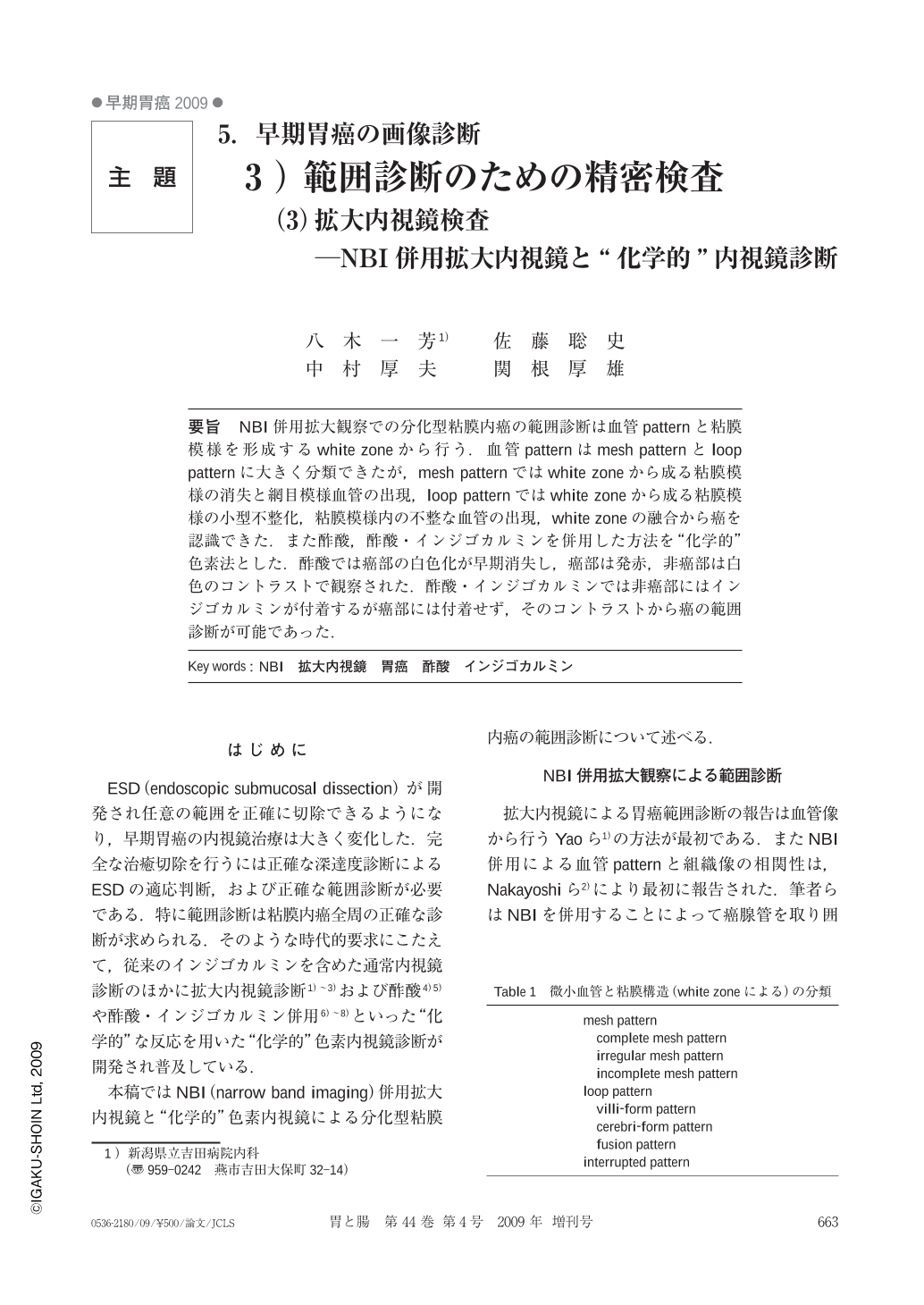
要旨 NBI併用拡大観察での分化型粘膜内癌の範囲診断は血管patternと粘膜模様を形成するwhite zoneから行う.血管patternはmesh patternとloop patternに大きく分類できたが,mesh patternではwhite zoneから成る粘膜模様の消失と網目模様血管の出現,loop patternではwhite zoneから成る粘膜模様の小型不整化,粘膜模様内の不整な血管の出現,white zoneの融合から癌を認識できた.また酢酸,酢酸・インジゴカルミンを併用した方法を“化学的”色素法とした.酢酸では癌部の白色化が早期消失し,癌部は発赤,非癌部は白色のコントラストで観察された.酢酸・インジゴカルミンでは非癌部にはインジゴカルミンが付着するが癌部には付着せず,そのコントラストから癌の範囲診断が可能であった.
The vascular patterns of differentiated adenocarcinomas were classified as mesh pattern and loop pattern. In case of mesh pattern, when the white zone, which indicates loss of mucosal pattern and mesh-like vascular pattern is no longer appearent, the extent of the cancer can be diagnosed. In case of loop pattern, unclear white zone, irregular and smaller mucosal pattern and irregular vascular pattern are indicators for diagnosis of cancerous extent.
The methods using acetic acid and indigocarmine with acetic acid have been called by authors “chemical-chromoendoscopy”. By using acetic acid(dynamic chemical method), a cancerous lesion is observed as a red area and the non-cancerous mucosa is observed as a white area because whitening due to acetic acid disappears in the cancerous area. In using indigocarmine with acetic acid(indigocarmine-acetic acid sandwich method), the cancerous lesion is observed as an indigocarmine-free area and non-cancerous mucosa is seen as the area to which indigocarmine adheres.
Copyright © 2009, Igaku-Shoin Ltd. All rights reserved.