Japanese
English
- 有料閲覧
PPV購入案内
1,320円 (1,200円+税10%) こちらは、1468ページから1476ページの文献です。購入には医書.jp本会員登録が必要です。
会員登録完了後に改めて上記「購入サイトへ」を選択してください。
または「購入サイトへ」選択後に「購入ログイン」、「新規会員登録」の順に進んでください。
会員登録について詳しくはをご確認ください。
- Abstract 文献概要
- 1ページ目 Look Inside
- 参考文献 Reference
- サイト内被引用 Cited by
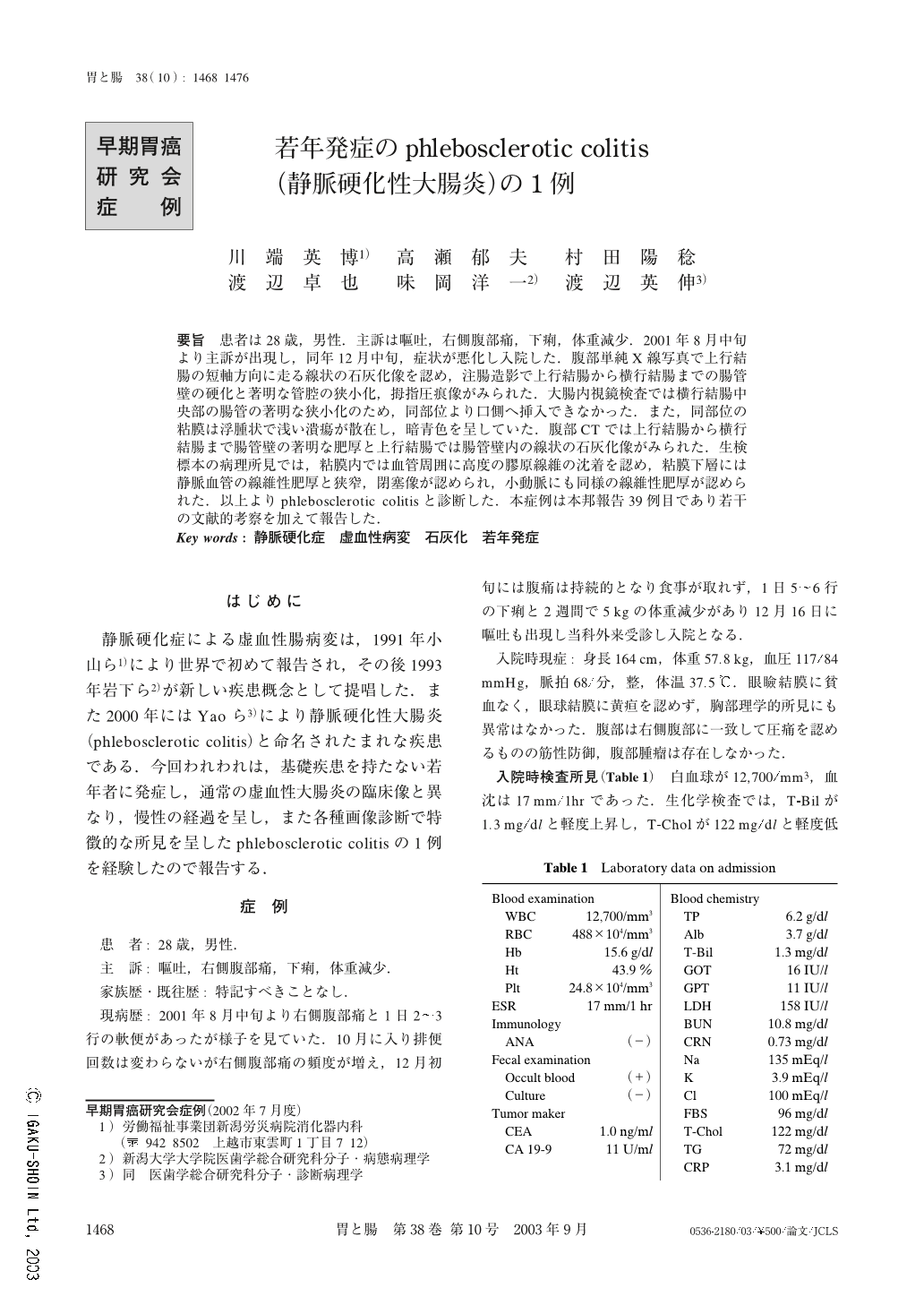
要旨 患者は28歳,男性.主訴は嘔吐,右側腹部痛,下痢,体重減少.2001年8月中旬より主訴が出現し,同年12月中旬,症状が悪化し入院した.腹部単純X線写真で上行結腸の短軸方向に走る線状の石灰化像を認め,注腸造影で上行結腸から横行結腸までの腸管壁の硬化と著明な管腔の狭小化,拇指圧痕像がみられた.大腸内視鏡検査では横行結腸中央部の腸管の著明な狭小化のため,同部位より口側へ挿入できなかった.また,同部位の粘膜は浮腫状で浅い潰瘍が散在し,暗青色を呈していた.腹部CTでは上行結腸から横行結腸まで腸管壁の著明な肥厚と上行結腸では腸管壁内の線状の石灰化像がみられた.生検標本の病理所見では,粘膜内では血管周囲に高度の膠原線維の沈着を認め,粘膜下層には静脈血管の線維性肥厚と狭窄,閉塞像が認められ,小動脈にも同様の線維性肥厚が認められた.以上よりphlebosclerotic colitisと診断した.本症例は本邦報告39例目であり若干の文献的考察を加えて報告した.
A 28-year-old man presented with right abdominal pain during the 4 months before his admission to the Labour Welfare Corporation Niigata Rosai Hospital. Abdominal conventional radiography showed a linear calcification of the vascular wall of the ascending colon. Barium enema examination showed a thumb-printing sign, sclerosis of the colonic wall and luminal narrowing of the ascending colon and transverse colon. Colonoscopic examination demonstrated dark blue mucosa, luminal narrowing and several small ulcers. Abdominal computed tomography revealed the thickening of the colonic wall and marked linear calcification of the vascular wall of the ascending colon and transverse colon. Based on these findings, the patient was diagnosed as having phlebosclerotic colitis. Phlebosclerotic colitis has rarely been reported. We discuss the clinical characteristics of this case.
Copyright © 2003, Igaku-Shoin Ltd. All rights reserved.