Japanese
English
- 有料閲覧
- Abstract 文献概要
- 1ページ目 Look Inside
- 参考文献 Reference
- サイト内被引用 Cited by
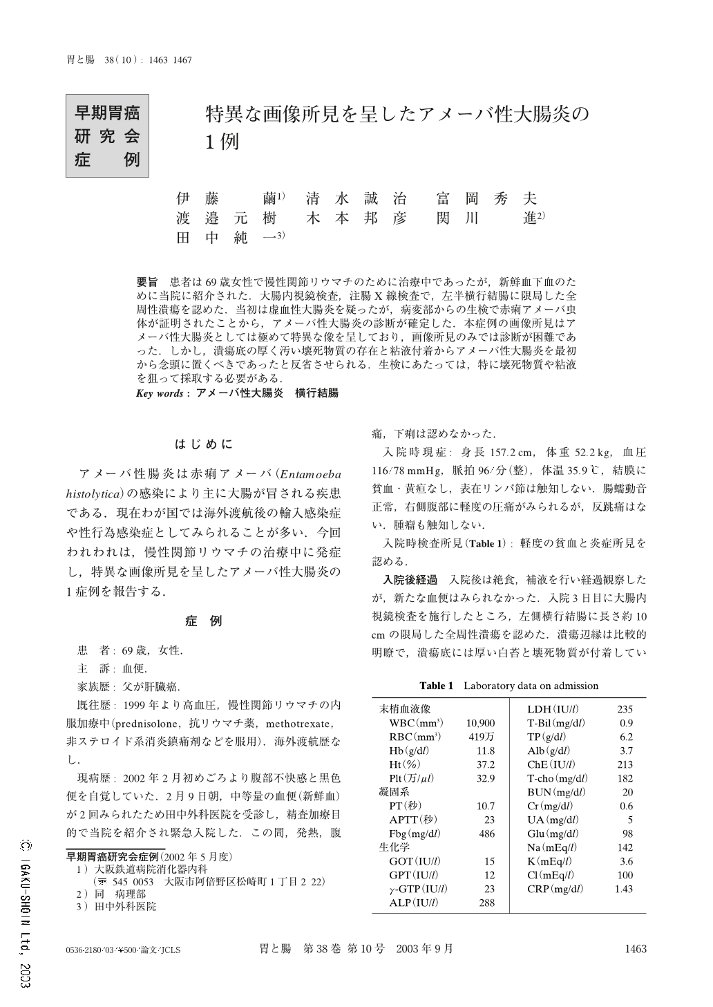
要旨 患者は69歳女性で慢性関節リウマチのために治療中であったが,新鮮血下血のために当院に紹介された.大腸内視鏡検査,注腸X線検査で,左半横行結腸に限局した全周性潰瘍を認めた.当初は虚血性大腸炎を疑ったが,病変部からの生検で赤痢アメーバ虫体が証明されたことから,アメーバ性大腸炎の診断が確定した.本症例の画像所見はアメーバ性大腸炎としては極めて特異な像を呈しており,画像所見のみでは診断が困難であった.しかし,潰瘍底の厚く汚い壊死物質の存在と粘液付着からアメーバ性大腸炎を最初から念頭に置くべきであったと反省させられる.生検にあたっては,特に壊死物質や粘液を狙って採取する必要がある.
A 69-year-old woman was referred to our hospital because of hematochezia. She had had a history of rheumatoid arthritis for four years, and had been treated with prednisolone, methotrexate, anti-rheumatoid arthritis drug, NSAID, and so on. Colonoscopy and barium enema X-ray revealed a circular ulcer with a length of about 10cm in the left side of the transverse colon. Although ischemic colitis was suspected from the findings at first, the demonstration of Entamoeba histolytica in one of three biopsy specimens established the diagnosis of amoebic colitis. The endoscopic and radiological findings of this case were so unusual that the diagnosis of amoebic colitis was difficult to make. However, thick and dirty necrotic substance and adhesion of thick mucus in the ulcer bottom were findings that are consistent with amoebic colitis. Biopsy should be taken from such regions to avoid misdiagnosis.

Copyright © 2003, Igaku-Shoin Ltd. All rights reserved.


