Japanese
English
- 有料閲覧
- Abstract 文献概要
- 1ページ目 Look Inside
- サイト内被引用 Cited by
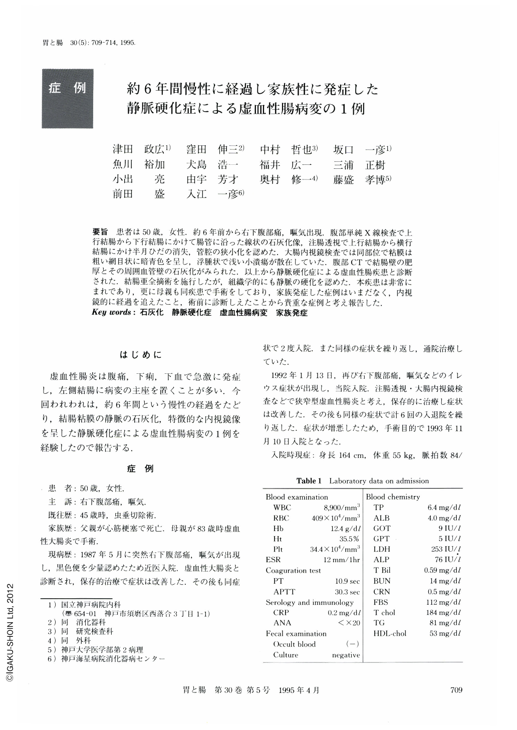
要旨 患者は50歳,女性.約6年前から右下腹部痛,嘔気出現.腹部単純X線検査で上行結腸から下行結腸にかけて腸管に沿った線状の石灰化像,注腸透視で上行結腸から横行結腸にかけ半月ひだの消失,管腔の狭小化を認めた.大腸内視鏡検査では同部位で粘膜は粗い網目状に暗青色を呈し,浮腫状で浅い小潰瘍が散在していた.腹部CTで結腸壁の肥厚とその周囲血管壁の石灰化がみられた.以上から静脈硬化症による虚血性腸疾患と診断された.結腸亜全摘術を施行したが,組織学的にも静脈の硬化を認めた.本疾患は非常にまれであり,更に母親も同疾患で手術をしており,家族発症した症例はいまだなく,内視鏡的に経過を追えたこと,術前に診断しえたことから貴重な症例と考え報告した.
A 50-year-old woman experienced right abdominal pain and nausea in the six years previous to her admission to the Kobe National Hospital. A plain abdominal x-ray film showed linear calcification of the vascular wall from the ascending colon to the discending colon. Barium enema showed disappearance of the semilunar fold, sclerosis of the colonic wall and luminal narrowing of the ascending colon and transverse colon. Colonoscopic examination showed an edematous and discolored (dark blue) mucosa, luminal narrowing and several small round ulcers. Abdominal CT examination showed the thickening of the colonic wall and marked tubular calcification of the vascular wall of the ascending colon and transverse colon. Based on these findings, the patient was diagnosed as having an ischemic intestinal lesion caused by phlebosclerosis. Because her severe symptoms repeated, she underwent subtotal colectomy. This disease has rarely been reported and we were able to diagnose it in this case before colectomy.

Copyright © 1995, Igaku-Shoin Ltd. All rights reserved.


