Japanese
English
- 有料閲覧
- Abstract 文献概要
- 1ページ目 Look Inside
- 参考文献 Reference
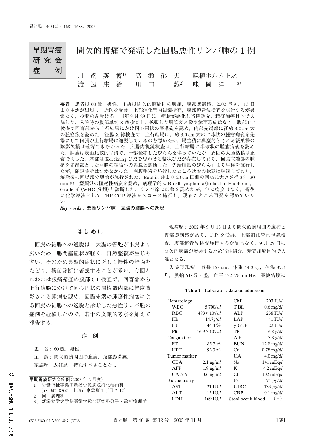
要旨 患者は60歳,男性.主訴は間欠的臍周囲の腹痛,腹部膨満感.2002年9月13日より主訴が出現し,近医を受診.上部消化管内視鏡検査,腹部超音波検査を試行するが異常なく,投薬のみ受ける.同年9月29日に,症状が悪化し当院紹介,精査加療目的で入院した.入院時の腹部単純X線検査上,拡張した腸管ガス像や鏡面形成はなく,腹部CT検査で回盲部から上行結腸にかけ同心円状の層構造を認め,内部先端部に径約3.0cm大の腫瘤像を認めた.注腸X線検査で,上行結腸に,約3.0cm大の半球状の腫瘤病変を先端にして回腸が上行結腸に逸脱しているのを認めたが,腸重積に典型的とされる蟹爪様の陰影欠損は確認できなかった.大腸内視鏡検査は,上行結腸に半球状の腫瘤病変を認めた.腫瘤は表面比較的平滑で,一部発赤したびらんを伴っていたが,周囲の大腸粘膜は正常であった.基部はKerckringひだを思わせる輪状ひだが存在しており,回腸末端部の腫瘍を先端部とした回腸の結腸への逸脱と診断した.先端腫瘍のびらん面より生検を施行したが,確定診断はつかなかった.開腹手術を施行したところ逸脱の状態は継続しており,解除後に回腸部分切除が施行された.Bauhin弁より20cm口側の回腸に大きさ径35×30mmの1型類似の隆起性病変を認め,病理学的にB-cell lymphoma(follicular lymphoma,Grade3)(WHO分類)と診断した.リンパ節に転移を認めたが,他に病変はなく,術後に化学療法としてTHP-COP療法を3コース施行し,現在のところ再発を認めていない.
A 60-year-old male was admitted with intermittent abdominal pain, two weeks prior to examination. Computerized tomography showed a concentric layered structure in the dilated colon. Subsequent Barium enema revealed a mass in the ascending colon, and colonoscopy disclosed a tumor measuring 3cm in diameter which prolapsed into the ascending colon from the small intestine. Preoperatively, primary malignant lymphoma of the terminal ileum was strongly suspected. Partial ileectomy was performed with lymph node dissection. The resected tumor was 35×30mm in size macroscopically. Histopathological diagnosis was a Mature B-cell neoplasm follicular lymphoma, Grade 3 according to WHO classification. Following the operation, chemotherapy was administered with THP-COP, and the patient has survived for 11 months without recurrence.

Copyright © 2005, Igaku-Shoin Ltd. All rights reserved.


