Japanese
English
- 有料閲覧
PPV購入案内
1,320円 (1,200円+税10%) こちらは、1785ページから1789ページの文献です。購入には医書.jp本会員登録が必要です。
会員登録完了後に改めて上記「購入サイトへ」を選択してください。
または「購入サイトへ」選択後に「購入ログイン」、「新規会員登録」の順に進んでください。
会員登録について詳しくはをご確認ください。
- Abstract 文献概要
- 1ページ目 Look Inside
- 参考文献 Reference
- サイト内被引用 Cited by
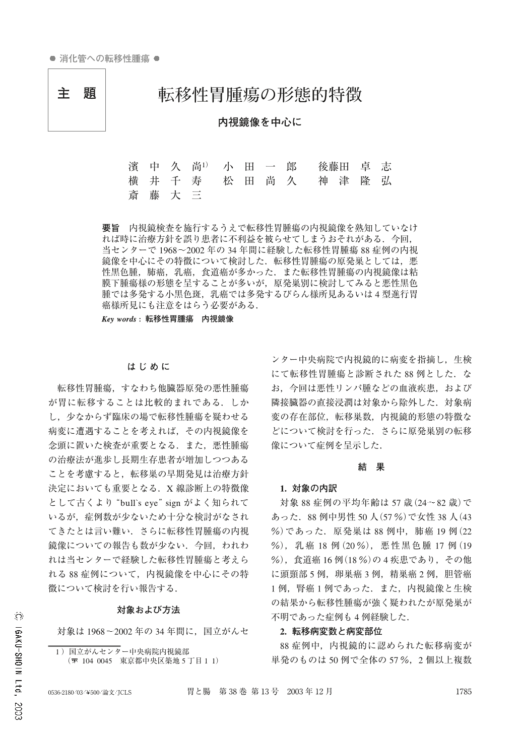
要旨 内視鏡検査を施行するうえで転移性胃腫瘍の内視鏡像を熟知していなければ時に治療方針を誤り患者に不利益を被らせてしまうおそれがある.今回,当センターで1968~2002年の34年間に経験した転移性胃腫瘍88症例の内視鏡像を中心にその特徴について検討した.転移性胃腫瘍の原発巣としては,悪性黒色腫,肺癌,乳癌,食道癌が多かった.また転移性胃腫瘍の内視鏡像は粘膜下腫瘍様の形態を呈することが多いが,原発巣別に検討してみると悪性黒色腫では多発する小黒色斑,乳癌では多発するびらん様所見あるいは4型進行胃癌様所見にも注意をはらう必要がある.
At the National Cancer Center Hospital, in the period from the 1968 to 2002, 88 cases of metastatic tumors of the stomach were detected and diagnosed by endoscopic examination and biopsy. In our study, primary cancers were frequently found in lung (22%), breast (20%), malignant melanoma (19%) and esophagus (18%).
Multiple lesions were seen in only 43% of these cases ; solitary metastases were more common than multiple metastases. Most of the metastatic tumors were located in the middle and upper gastric body, mainly on the greater curvature. Endoscopic appearance was broadly classified into 3 categories, which were (1) submucosal tumor-like formation (50%), (2) cancer-like formation (33%) and (3) others (17%). In category (1), 70% of them had a central depression. Concerning category (2), 23 out of 29 cases resembled advanced cancer. In conclusion, it is necessary to have a better understanding of the appearance of metastatic tumors of the stomach, so we analyzed their endoscopic features. Endoscopic examination is helpful both for detecting tumors and for indicating their proper treatment.
Copyright © 2003, Igaku-Shoin Ltd. All rights reserved.