Japanese
English
- 有料閲覧
PPV購入案内
1,320円 (1,200円+税10%) こちらは、1251ページから1258ページの文献です。購入には医書.jp本会員登録が必要です。
会員登録完了後に改めて上記「購入サイトへ」を選択してください。
または「購入サイトへ」選択後に「購入ログイン」、「新規会員登録」の順に進んでください。
会員登録について詳しくはをご確認ください。
- Abstract 文献概要
- 1ページ目 Look Inside
- 参考文献 Reference
- サイト内被引用 Cited by
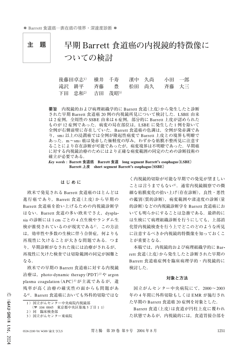
要旨 内視鏡的および病理組織学的にBarrett食道(上皮)から発生したと診断された早期Barrett食道癌20例の内視鏡所見について検討した.LSBE由来は2症例,全周性のSSBE由来は6症例,部分的にBarrett上皮が認められたものが12症例であった.病変の局在部位は,LSBEに発生した1例を除いて全例が右側前壁に存在していた.Barrett食道癌の色調は,全例が発赤調であり,sm2以上の浸潤癌では全例が隆起性病変でBarrett上皮との境界も明瞭であった.m~sm1癌は発赤した極軽度の厚み,わずかな粘膜不整所見に注意することにより存在診断が可能であったが,病変境界は不明瞭であった.早期癌に対する内視鏡治療のためにはより正確な病変範囲の同定のための診断技術の確立が必要である.
Increasing incidence of Barrett's cancer has been reported in the West. However, almost all cases have been detected in the advanced stage. The endoscopic features of 20 superficial Barrett's cancers were evaluated endoscopically. Two Barrett's cancers had developed from LSBE, and 6 lesions had originated from circular SSBE. Barrett's epithelium on a part of the lower esophagus (non-circular SSBE) was found in 12 cases.
Almost all cases except for 1 case developed from LSBE had a superficial tumor on the right aspect of the lower esophagus. All lesions could be detected endoscopically by their reddish mucosal changes. In particular, submucosal invasive cancers were easily detected due to their elevated component. However, mucosal lesions revealed such findings as slight thickness or subtle irregularity, which were less indicative of malignancy. Further investigations using larger numbers of lesions or development of new endoscopic procedures should be considered for the detection of Barrett's cancer at an early stage.
1) Endoscopy Division, National Cancer Center Hospital, Tokyo
Copyright © 2004, Igaku-Shoin Ltd. All rights reserved.