Japanese
English
- 有料閲覧
PPV購入案内
1,320円 (1,200円+税10%) こちらは、835ページから843ページの文献です。購入には医書.jp本会員登録が必要です。
会員登録完了後に改めて上記「購入サイトへ」を選択してください。
または「購入サイトへ」選択後に「購入ログイン」、「新規会員登録」の順に進んでください。
会員登録について詳しくはをご確認ください。
- Abstract 文献概要
- 1ページ目 Look Inside
- 参考文献 Reference
- サイト内被引用 Cited by
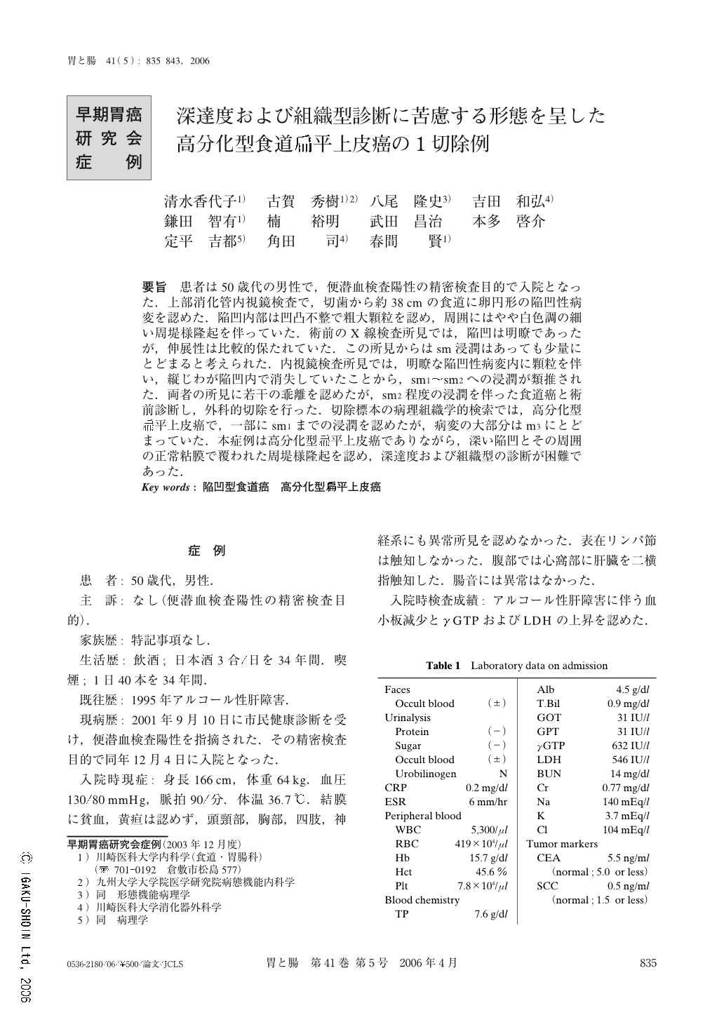
要旨 患者は50歳代の男性で,便潜血検査陽性の精密検査目的で入院となった.上部消化管内視鏡検査で,切歯から約38cmの食道に卵円形の陥凹性病変を認めた.陥凹内部は凹凸不整で粗大顆粒を認め,周囲にはやや白色調の細い周堤様隆起を伴っていた.術前のX線検査所見では,陥凹は明瞭であったが,伸展性は比較的保たれていた.この所見からはsm浸潤はあっても少量にとどまると考えられた.内視鏡検査所見では,明瞭な陥凹性病変内に顆粒を伴い,縦じわが陥凹内で消失していたことから,sm1~sm2への浸潤が類推された.両者の所見に若干の乖離を認めたが,sm2程度の浸潤を伴った食道癌と術前診断し,外科的切除を行った.切除標本の病理組織学的検索では,高分化型扁平上皮癌で,一部にsm1までの浸潤を認めたが,病変の大部分はm3にとどまっていた.本症例は高分化型扁平上皮癌でありながら,深い陥凹とその周囲の正常粘膜で覆われた周堤様隆起を認め,深達度および組織型の診断が困難であった.
A 50-year-old Japanese man was admitted to our hospital because of a positive fecal occult blood test result.
Esophagoscopy revealed a crater-like, oval depressed lesion with an undulated surface in the lower thoracic esophagus. Around the depression, a narrow protrusion covered with normal mucosa was recognized. The lesion was unstained with iodine. An endoscopic diagnosis of esophageal cancer invading the submucosal layer was made. Surprisingly, a barium examination disclosed the wall rigidity of the lesion to be extremely mild despite an evident depression followed by a rim-like surrounding elevation. Based on these findings, an unusual esophageal neoplasm, such as a poorly differentiated squamous cell carcinoma was suspected.
Macroscopic examination of the resected specimen disclosed a whitish oval depression measuring 13×8 mm in size. Microscopically, the tumor consisted of well differentiated squamous cell carcinoma, which was minimally invading the submucosal layer. Marked lymphocytic infiltration was also seen.
In this paper, we describe in detail an endoscopically, radiologically and histopathologically interesting case of esophageal cancer.
Copyright © 2006, Igaku-Shoin Ltd. All rights reserved.