Japanese
English
- 有料閲覧
PPV購入案内
2,200円 (2,000円+税10%) こちらは、971ページから977ページの文献です。購入には医書.jp本会員登録が必要です。
会員登録完了後に改めて上記「購入サイトへ」を選択してください。
または「購入サイトへ」選択後に「購入ログイン」、「新規会員登録」の順に進んでください。
会員登録について詳しくはをご確認ください。
- Abstract 文献概要
- 1ページ目 Look Inside
- 参考文献 Reference
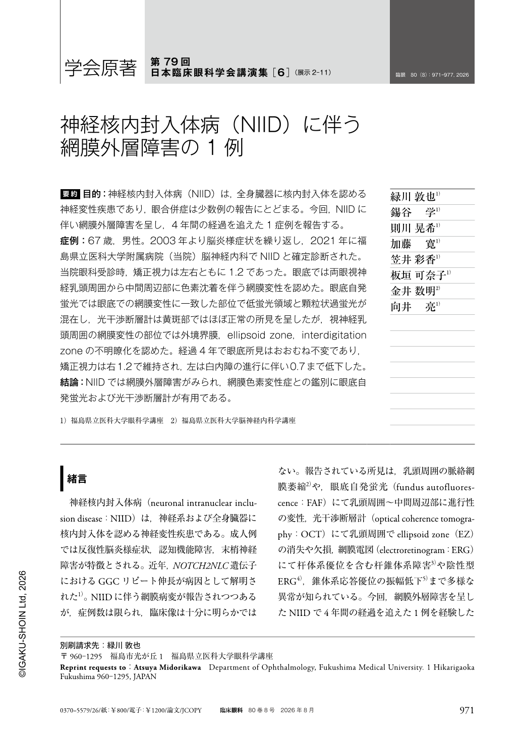
要約 目的:神経核内封入体病(NIID)は,全身臓器に核内封入体を認める神経変性疾患であり,眼合併症は少数例の報告にとどまる。今回,NIIDに伴い網膜外層障害を呈し,4年間の経過を追えた1症例を報告する。
症例:67歳,男性。2003年より脳炎様症状を繰り返し,2021年に福島県立医科大学附属病院(当院)脳神経内科でNIIDと確定診断された。当院眼科受診時,矯正視力は左右ともに1.2であった。眼底では両眼視神経乳頭周囲から中間周辺部に色素沈着を伴う網膜変性を認めた。眼底自発蛍光では眼底での網膜変性に一致した部位で低蛍光領域と顆粒状過蛍光が混在し,光干渉断層計は黄斑部ではほぼ正常の所見を呈したが,視神経乳頭周囲の網膜変性の部位では外境界膜,ellipsoid zone,interdigitation zoneの不明瞭化を認めた。経過4年で眼底所見はおおむね不変であり,矯正視力は右1.2で維持され,左は白内障の進行に伴い0.7まで低下した。
結論:NIIDでは網膜外層障害がみられ,網膜色素変性症との鑑別に眼底自発蛍光および光干渉断層計が有用である。
Abstract Purpose:To report a long-term prognosis of neuronal intranuclear inclusion disease(NIID) presenting with outer retinal degeneration.
Case:A 67-year-old man with a history of recurrent encephalopathy since 2003 was diagnosed with NIID at our institution in 2021. At the initial ophthalmic examination, best-corrected visual acuity was 1.2 in both eyes. Fundus examination revealed retinal degeneration with bilateral pigmentary changes extending from the peripapillary region to the midperiphery. Fundus autofluorescence showed areas of hypo-autofluorescence corresponding to degenerative lesions intermingled with granular hyper-autofluorescence. Optical coherence tomography revealed an almost normal retinal architecture in the macular area. However, disruption of the external limiting membrane, ellipsoid zone, and interdigitation zone was observed in the peripapillary region. Fundus findings remained largely unchanged over the 4-year follow-up period. Visual acuity was maintained at 1.2 in the right eye, while that in the left eye decreased to 0.7 owing to cataract progression.
Conclusion:Outer retinal degeneration can be associated with NIID. Fundus autofluorescence and optical coherence tomography are useful in distinguishing this condition from retinitis pigmentosa. Comprehensive ophthalmic evaluation should be considered in patients with NIID.
Copyright © 2026, Igaku-Shoin Ltd. All rights reserved.