Japanese
English
- 有料閲覧
PPV購入案内
1,320円 (1,200円+税10%) こちらは、248ページから254ページの文献です。購入には医書.jp本会員登録が必要です。
会員登録完了後に改めて上記「購入サイトへ」を選択してください。
または「購入サイトへ」選択後に「購入ログイン」、「新規会員登録」の順に進んでください。
会員登録について詳しくはをご確認ください。
- Abstract 文献概要
- 1ページ目 Look Inside
- 参考文献 Reference
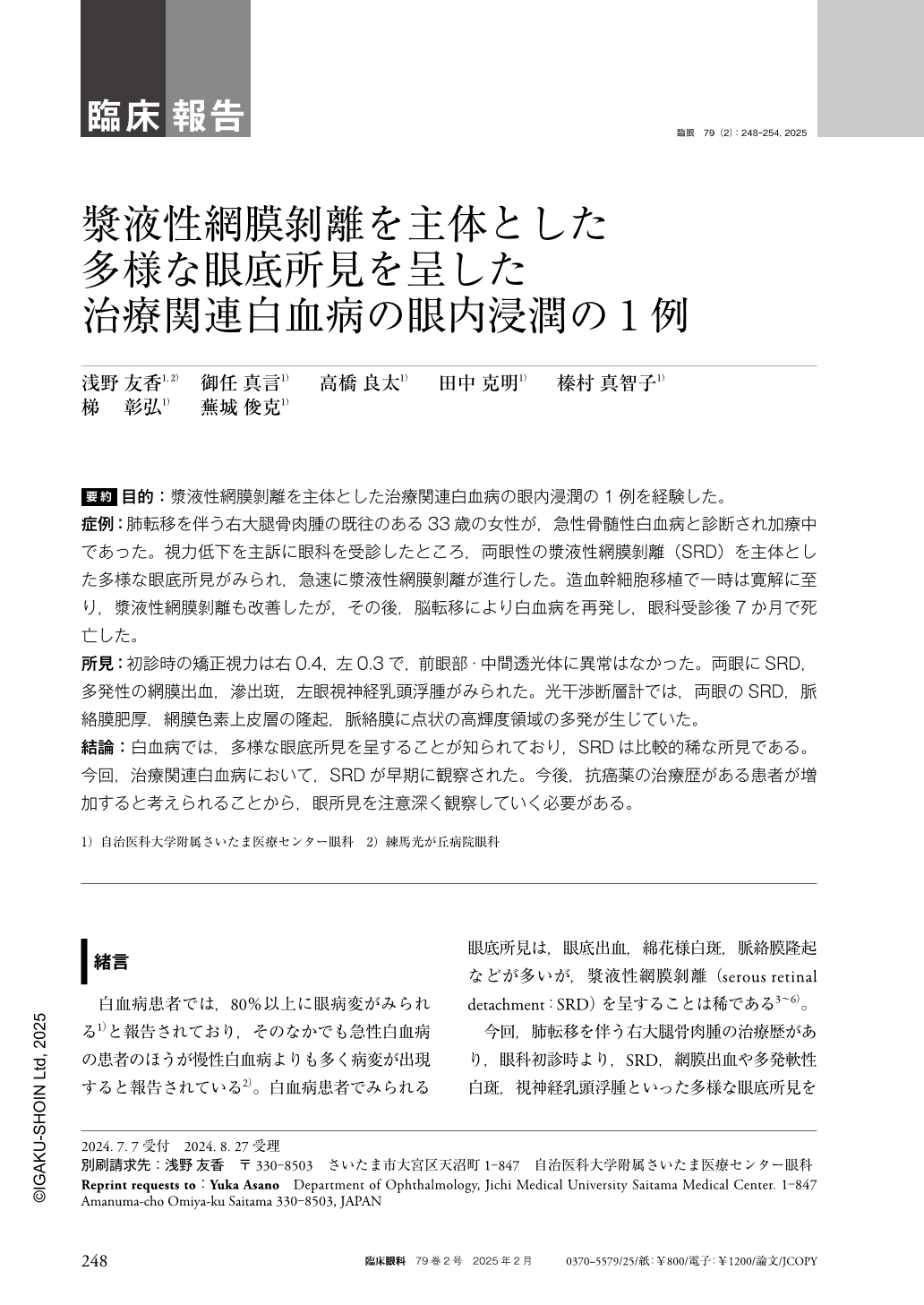
要約 目的:漿液性網膜剝離を主体とした治療関連白血病の眼内浸潤の1例を経験した。
症例:肺転移を伴う右大腿骨肉腫の既往のある33歳の女性が,急性骨髄性白血病と診断され加療中であった。視力低下を主訴に眼科を受診したところ,両眼性の漿液性網膜剝離(SRD)を主体とした多様な眼底所見がみられ,急速に漿液性網膜剝離が進行した。造血幹細胞移植で一時は寛解に至り,漿液性網膜剝離も改善したが,その後,脳転移により白血病を再発し,眼科受診後7か月で死亡した。
所見:初診時の矯正視力は右0.4,左0.3で,前眼部・中間透光体に異常はなかった。両眼にSRD,多発性の網膜出血,滲出斑,左眼視神経乳頭浮腫がみられた。光干渉断層計では,両眼のSRD,脈絡膜肥厚,網膜色素上皮層の隆起,脈絡膜に点状の高輝度領域の多発が生じていた。
結論:白血病では,多様な眼底所見を呈することが知られており,SRDは比較的稀な所見である。今回,治療関連白血病において,SRDが早期に観察された。今後,抗癌薬の治療歴がある患者が増加すると考えられることから,眼所見を注意深く観察していく必要がある。
Abstract Purpose:We report a case of intraocular infiltration in therapy-related leukemia, primarily associated with serous retinal detachment.
Case:A 33-year-old female with a history of osteosarcoma of the right femur with pulmonary metastases was diagnosed with acute myeloid leukemia(AML)and was undergoing treatment. During the course, she presented to the ophthalmology department with visual impairment. Various fundus abnormalities, predominantly serous retinal detachment, were observed in both eyes. However, serous retinal detachment progressed rapidly. Temporary remission was achieved via hematopoietic stem cell transplantation, which improved the serous retinal detachment. Subsequently, leukemia relapsed because of brain metastasis, and she died seven months following the ophthalmology consultation.
Findings:The initial corrected visual acuity was 0.4 right and 0.3 left, with no abnormalities in the anterior segment or vitreous. Fundus examination revealed serous retinal detachment, retinal hemorrhages, and exudates in both eyes. The left eye exhibited papilledema. Optical coherence tomography revealed serous retinal detachment in both eyes, choroidal thickening, bumps in the retinal pigment epithelium, and multiple bright spots in the choroid.
Conclusion:We report a case of therapy-related leukemia with early-onset serous retinal detachment and poor prognosis. Leukemia is known to present with a variety of fundus findings, among which serous retinal detachment is a relatively rare finding. Since the number of patients who have been treated with anticancer agents is expected to increase in the future, we need to carefully monitor the ocular findings.
Copyright © 2025, Igaku-Shoin Ltd. All rights reserved.