Japanese
English
- 有料閲覧
- Abstract 文献概要
- 1ページ目 Look Inside
- 参考文献 Reference
はじめに
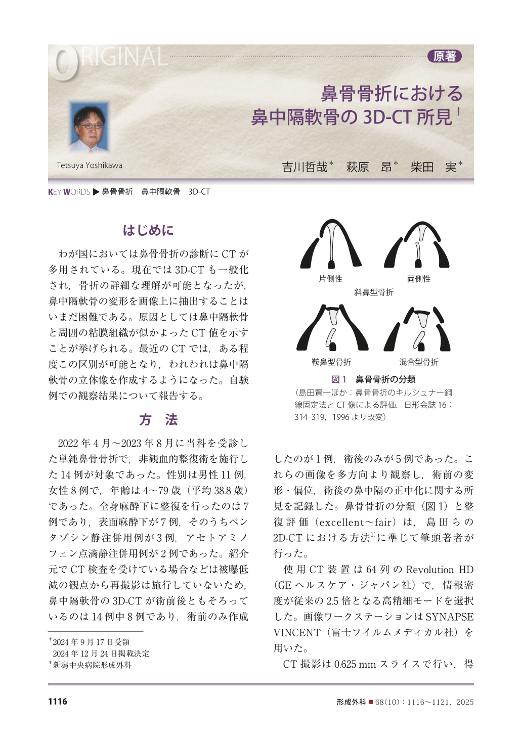
わが国においては鼻骨骨折の診断にCTが多用されている。現在では3D-CTも一般化され,骨折の詳細な理解が可能となったが,鼻中隔軟骨の変形を画像上に抽出することはいまだ困難である。原因としては鼻中隔軟骨と周囲の粘膜組織が似かよったCT値を示すことが挙げられる。最近のCTでは,ある程度この区別が可能となり,われわれは鼻中隔軟骨の立体像を作成するようになった。自験例での観察結果について報告する。
In cases involving nasal bone fractures, it has been difficult to visualize the nasal septal cartilage by conventional CT, thereby limiting the precise understanding of deformities of the nasal septal cartilage based on imaging. With recent advances in CT technology, the creation of 3D-CT models of the septal cartilage has become possible (although not optimal). We report our observations and experience with such models. With the use of a high-resolution CT Revolution HD Ⓡ system and Synapse Vincent Ⓡ software, we created 3D-CT models of the septal cartilage for the 14 cases of acute nasal fracture treated in our department since April 2022. The patientsʼ preoperative findings included mainly deviation of the upper septal cartilage, displacement to the opposite side, and various irregularities. Postoperatively, straightening of the septal cartilage was observed in almost all of the cases. We observed that by visualizing the deformity of the septal cartilage in three dimensions, the pathology of nasal bone fractures can be clarified. The creation of 3D-CT models of the septal cartilage has the advantages of (i) enabling the sharing of the images with other clinicians and (ii) providing a visual record of the deformities. Although the clarity of these models remains sub-optimal, techniques such as color-coding the left and right surfaces of the cartilage and reducing the opacity of bones have facilitated a better understanding of deformities of the nasal septal cartilage.
Copyright© 2025 KOKUSEIDO CO., LTD. All Rights Reserved.