Japanese
English
- 有料閲覧
- Abstract 文献概要
- 1ページ目 Look Inside
- 参考文献 Reference
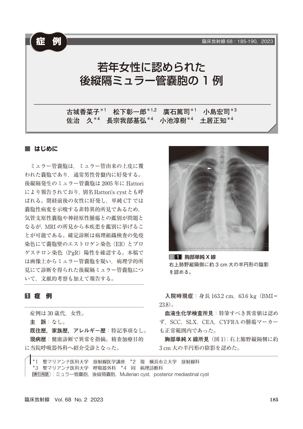
ミュラー管嚢胞は,ミュラー管由来の上皮に覆われた嚢胞であり,通常男性骨盤内に好発する。後縦隔発生のミュラー管嚢胞は2005年にHattoriにより報告されており,別名Hattori’s cystとも呼ばれる。閉経前後の女性に好発し,単純CTでは嚢胞性病変を示唆する非特異的所見であるため,気管支原性嚢胞や神経原性腫瘍との鑑別が問題となるが,MRIの所見から本疾患を鑑別に挙げることが可能である。確定診断は病理組織検査の免疫染色にて嚢胞壁のエストロゲン染色(ER)とプロゲステロン染色(PgR)陽性を確認する。本稿では画像上からミュラー管嚢胞を疑い,病理学的所見にて診断を得られた後縦隔ミュラー管嚢胞について,文献的考察も加えて報告する。
A 30-year-old woman in her 30 s was noted to have an abnormal shadow on a plain chest photograph during a physical examination. A plain chest X-ray showed a half-round mass shadow on the mediastinal side of the right upper lung field. A plain CT scan of the chest showed a well-defined cystic mass of 23×23×30 mm in size on the right side of the Th3 vertebral body. A plain MRI showed a water density signal and no internal substantial component. There was also no continuity between the cyst and the spinal canal. Based on the images, Müllerian duct cyst was most suspected, and surgery was performed. Histopathology revealed a cyst wall structure lined by a single layer of columnar epithelium. Immunostaining was positive for estrogen receptor(ER)and progesterone receptor(PgR), and a diagnosis of Müllerian duct cyst was made. Müllerian duct cysts arising in the posterior mediastinum can be differentiated from bronchogenic or neurogenic cysts by their site of origin. The definitive diagnosis is made by histopathology, which confirms ER and PgR positivity of the cyst wall.

Copyright © 2023, KANEHARA SHUPPAN Co.LTD. All rights reserved.


