Japanese
English
症例
Robinow症候群の2例
Two cases of Robinow syndrome
米田 昌平
1
,
下野 太郎
1
,
藤田 賢司
1
,
立川 裕之
1
,
瀬戸 俊之
2
,
三木 幸雄
1
Shohei Yoneda
1
1大阪市立大学大学院医学研究科 放射線診断学・IVR学
2同 発達小児医学分野
1Department of Diagnostic and Interventional Radiology Osaka City University Graduate School of Medicine
キーワード:
メッケル腟拡大
,
骨軟骨形成異常
,
脳梁・脳幹の軽度萎縮
Keyword:
メッケル腟拡大
,
骨軟骨形成異常
,
脳梁・脳幹の軽度萎縮
pp.833-838
発行日 2018年7月10日
Published Date 2018/7/10
DOI https://doi.org/10.18888/rp.0000000492
- 有料閲覧
- Abstract 文献概要
- 1ページ目 Look Inside
- 参考文献 Reference
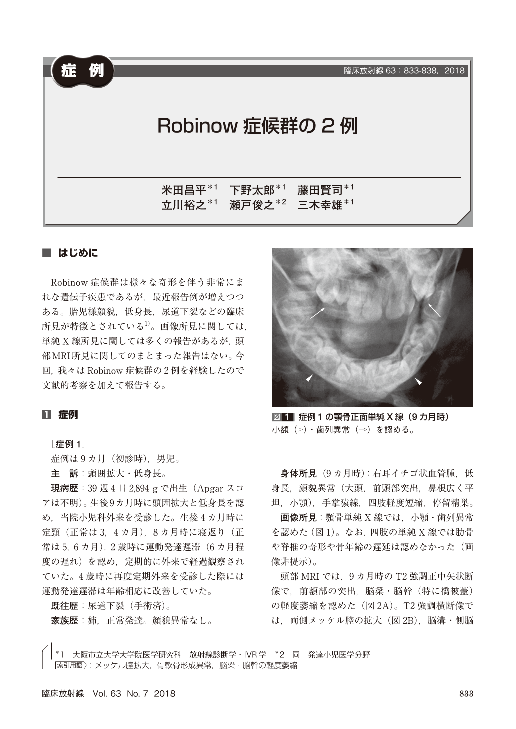
Robinow症候群は様々な奇形を伴う非常にまれな遺伝子疾患であるが,最近報告例が増えつつある。胎児様顔貌,低身長,尿道下裂などの臨床所見が特徴とされている1)。画像所見に関しては,単純X線所見に関しては多くの報告があるが,頭部MRI所見に関してのまとまった報告はない。今回,我々はRobinow症候群の2例を経験したので文献的考察を加えて報告する。
We report two children of Robinow syndrome, a rare disease that affects the development of many parts of the body, particularly in bones. On brain MRI, both cases showed mild atrophy of the corpus callosum and brainstem, and mild enlargement of the lateral ventricles. In addition, the first case showed enlargement of Meckel’s cave, which may be caused by remodeling of the adjacent petrous temporal bone. These imaging findings may be characteristics of Robinow syndrome.

Copyright © 2018, KANEHARA SHUPPAN Co.LTD. All rights reserved.


