Japanese
English
- 有料閲覧
- Abstract 文献概要
- 1ページ目 Look Inside
- 参考文献 Reference
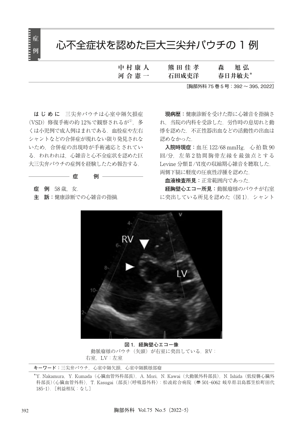
はじめに 三尖弁パウチは心室中隔欠損症(VSD)修復手術の約12%で観察されるが1),多くは小児例で成人例はまれである.血栓症や左右シャントなどの合併症が現れない限り発見されないため,合併症の出現時が手術適応とされている.われわれは,心雑音と心不全症状を認めた巨大三尖弁パウチの症例を経験したため報告する.
Tricuspid pouch forms during the spontaneous closure of a ventricular septal defect (VSD). Cases have been reported in which the tricuspid pouch was discovered for the first time during surgery and could not be distinguished from an aneurysm of the membranous septum (AMS). A 58-year-old woman had a heart murmur. Transthoracic echocardiography showed an aneurysm-like pouch protruding into the right ventricle. Magnetic resonance imaging could not distinguish between AMS and tricuspid pouch;however, contrast-enhanced computed tomography showed a VSD. The membranous structure comprised multiple lobules, and the tendon of the papillary muscles was continuous with the tricuspid valve. Intraoperatively, the tricuspid valve septal leaflet was adhered to the defect hole. It was incised along the annulus, the VSD was closed with a bovine pericardial patch, and the annulus of the tricuspid valve septal leaflet was suture closed. The patient was discharged after a good postoperative course.

© Nankodo Co., Ltd., 2022


