Japanese
English
- 有料閲覧
- Abstract 文献概要
- 1ページ目 Look Inside
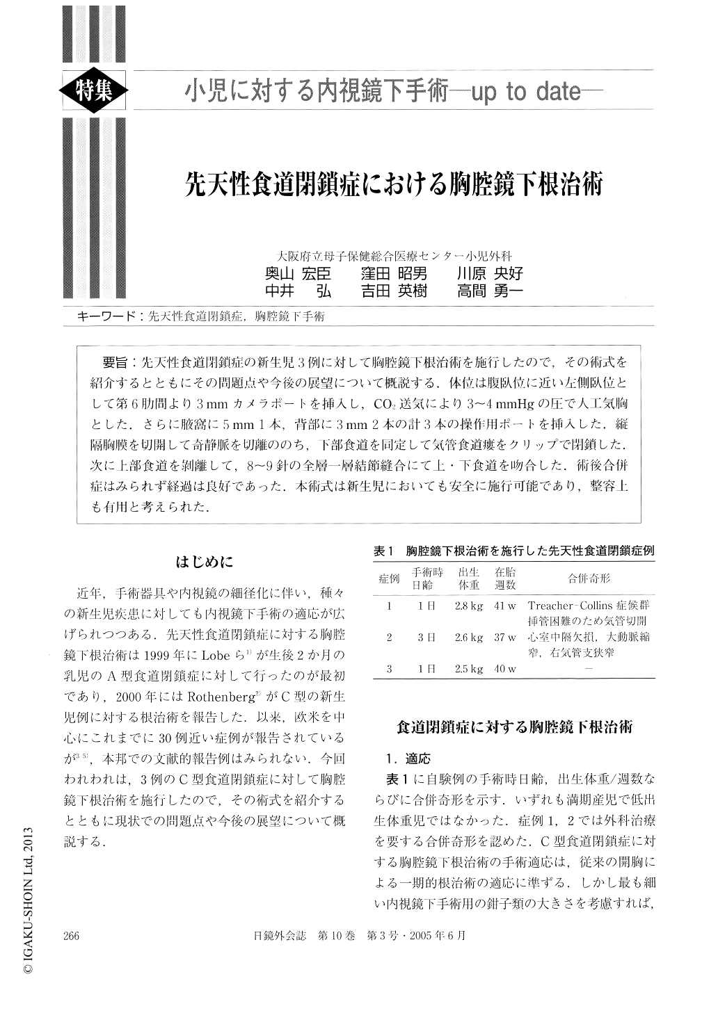
先天性食道閉鎖症の新生児3例に対して胸腔鏡下根治術を施行したので,その術式を紹介するとともにその問題点や今後の展望について概説する.体位は腹臥位に近い左側臥位として第6肋間より3mmカメラポートを挿入し,CO2送気により3〜4mmHgの圧で人工気胸とした.さらに腋窩に5mm 1本,背部に3mm 2本の計3本の操作用ポートを挿入した.縦隔胸膜を切開して奇静脈を切離ののち,下部食道を同定して気管食道瘻をクリップで閉鎖した.次に上部食道を?離して,8〜9針の全層一層結節縫合にて上・下食道を吻合した.術後合併症はみられず経過は良好であった.本術式は新生児においても安全に施行可能であり,整容上も有用と考えられた.
We present three neonates with esophageal atresia and distal tracheoesophageal fistula (TEF) treated with primary thoracoscopic repair. The patients were positioned prone with the right side slightly elevated. Firstly, a 3 mm camera port was inserted in the sixth intercostal space, and the pleural space was insufflated to a pres-sure of 3 to 4 mmHg. Additional 3 working ports (5 mm × 1.3 mm × 2) were placed in the axillary and back re-gion. The pleura was incised and the azygos vein was divided with the ultrasonic coagulating shears.

Copyright © 2005, JAPAN SOCIETY FOR ENDOSCOPIC SURGERY All rights reserved.


